Co-reporter: Paweł M. Antonik, Alexander N. Volkov, Ursula N. Broder, Daniele Lo Re, Nico A. J. van Nuland, and Peter B. Crowley
pp: 1195-1203
Publication Date(Web):February 4, 2016
DOI: 10.1021/acs.biochem.5b01212
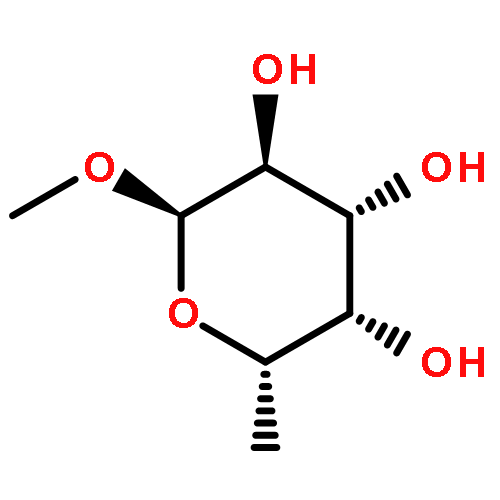
Sugar binding by a cell surface ∼29 kDa lectin (RSL) from the bacterium Ralstonia solanacearum was characterized by NMR spectroscopy. The complexes formed with four monosaccharides and four fucosides were studied. Complete resonance assignments and backbone dynamics were determined for RSL in the sugar-free form and when bound to l-fucose or d-mannose. RSL was found to interact with both the α- and the β-anomer of l-fucose and the “fucose like” sugars d-arabinose and l-galactose. Peak splitting was observed for some resonances of the binding site residues. The assignment of the split signals to the α- or β-anomer was confirmed by comparison with the spectra of RSL bound to methyl-α-l-fucoside or methyl-β-l-fucoside. The backbone dynamics of RSL were sensitive to the presence of ligand, with the protein adopting a more compact structure upon binding to l-fucose. Taking advantage of tryptophan residues in the binding sites, we show that the indole resonance is an excellent reporter on ligand binding. Each sugar resulted in a distinct signature of chemical shift perturbations, suggesting that tryptophan signals are a sufficient probe of sugar binding.
Co-reporter: Paweł M. Antonik, Ahmed M. Eissa, Adam R. Round, Neil R. Cameron, and Peter B. Crowley
pp:
Publication Date(Web):July 12, 2016
DOI: 10.1021/acs.biomac.6b00766
PEGylation, the covalent modification of proteins with polyethylene glycol, is an abundantly used technique to improve the pharmacokinetics of therapeutic proteins. The drawback with this methodology is that the covalently attached PEG can impede the biological activity (e.g., reduced receptor-binding capacity). Protein therapeutics with “disposable” PEG modifiers have potential advantages over the current technology. Here, we show that a protein–polymer “Medusa complex” is formed by the combination of a hexavalent lectin with a glycopolymer. Using NMR spectroscopy, small-angle X-ray scattering (SAXS), size exclusion chromatography, and native gel electrophoresis it was demonstrated that the fucose-binding lectin RSL and a fucose-capped polyethylene glycol (Fuc-PEG) form a multimeric assembly. All of the experimental methods provided evidence of noncovalent PEGylation with a concomitant increase in molecular mass and hydrodynamic radius. The affinity of the protein–polymer complex was determined by ITC and competition experiments to be in the micromolar range, suggesting that such systems have potential biomedical applications.
