Co-reporter:Meng Zhao, Shan-Shan Xue, Xue-Qin Jiang, Limin Zheng, Liang-Nian Ji, Zong-Wan Mao
Journal of Molecular Catalysis A: Chemical 2015 Volume 396() pp:346-352
Publication Date(Web):January 2015
DOI:10.1016/j.molcata.2014.10.020
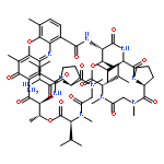
•A dinuclear Co(II) complex, Co2L, coupling to β-CDs was synthesized and characterized.•Supramolecular effect protects the dinuclear Co(II) center from oxidation in air.•High activity toward bis(4-nitrophenyl) phosphate hydrolysis was observed at neutral pH.•Metal effect was discussed by comparing the analogous Zn2L, Cu2L and Co2L.•Unexpected phosphotase-like activity was observed for Co2L.Dinuclear metallohydrolases are ubiquitously involved in biological transformations. For understanding and application purpose, significant efforts have been dedicated to the structural or/and functional mimics. Herein, a dinuclear Co(II) complex, Co2L, with intramolecular β-CDs is presented. The β-CDs confine a hydrophobic microenvironment that contributes to the valence stability of the ligated Co(II) ions. Species of Co2L in aqueous solution is clarified according to the results of potentiometric titration and UV–vis titration experiments. A highly positive-charged species [Co2HL]4+ is present at quasi-neutral condition, which contributed significantly to the binding and activity toward bis(4-nitrophenyl) phosphate (BNPP) assisted by the hydrophobic interactions exerted by intramolecular β-CDs. The kcat/KM value of Co2L is determined to be 9.8 × 10⁻² M−1 s−1 at pH 6.5 and 308 K, which exceeds that of the reported zinc analog (Zn2L) by three orders of magnitude. Unexpectedly, Co2L also catalyzes hydrolysis of a phosphate monoester, 4-nitrophenyl phosphate (NPP). The observed phosphate esterase activities are rarely reported among synthetic Co(II) complexes.A dinuclear Co(II) complex equipped with intramolecular β-CDs effectively catalyzed the hydrolysis of phosphate esters. Metal effect is discussed.
Co-reporter:Wen-Chao Wu;Hong-Wei Sun;Hai-Tian Chen;Jing Liang;Xing-Juan Yu;Chong Wu;Zilian Wang
PNAS 2014 111 (11 ) pp:4221-4226
Publication Date(Web):2014-03-18
DOI:10.1073/pnas.1320753111
Cancer is associated with a profound perturbation in myelopoiesis that results in the accumulation of myeloid-derived suppressor
cells (MDSCs) to promote disease progression. Recent studies in mice suggest that tumor-derived factors could regulate the
differentiation of hematopoietic stem and progenitor cells (HSPCs) in the bone marrow and subsequently contribute to dysregulation
of hematopoiesis. However, the nature and role of HPSCs in patients with cancer remain unknown. Here we show, in detailed
studies of the peripheral blood from 133 untreated patients with seven different types of tumors, that the composition of
circulating HSPCs was significantly altered in patients with solid tumors. The frequencies of circulating granulocyte–monocyte
progenitors (GMPs) were increased four to seven fold in all types of tumors examined, and the circulating hematopoietic precursors
exhibited myeloid bias with a skew toward granulocytic differentiation in patients with solid tumors. These myeloid precursors
are selectively enriched in tumor tissues, and the high levels of circulating GMPs were positively correlated with disease
progression. By using cord blood-derived CD34+ cells, we developed an in vitro short-term culture model to effectively induce the rapid generation of MDSCs. We found that,
among the factors produced by various tumors, GM-CSF, granulocyte colony-stimulating factor, and IL-6 could not only promote
the myeloid-biased differentiation, but also induce the differentiation of myeloid precursors into functional MDSCs. These
findings suggest that the altered circulating HSPCs may serve as an important link between dysregulated bone marrow hematopoiesis
and accumulated MDSCs in patients with cancer.
Co-reporter:Yuan Liao;Bo Wang;Jinqing Li;Lian Li;Xingjuan Yu;Jing Xu;Yi Zhang;Shupeng Chen;Huilan Rao;Lanjun Zhang;Limin Zheng
Cancer Immunology, Immunotherapy 2013 Volume 62( Issue 10) pp:
Publication Date(Web):2013/10/01
DOI:10.1007/s00262-013-1460-4
The proinflammatory cytokine interleukin 17 (IL-17) is considered to play a crucial role in diverse human tumors; however, its role in disease progression remains controversial. This study investigated the cellular source and distribution of IL-17 in esophageal squamous cell carcinoma (ESCC) in situ and determined its prognostic value. Immunohistochemistry, immunofluorescence and immunoelectron microscopy were used to identify IL-17-expressing cells in ESCC tissues, paying particular attention to their anatomic localization. Kaplan–Meier analysis and Cox proportional hazards regression models were applied to estimate overall survival in 215 ESCC patients with long-term follow-up (>10 years). The results showed that mast cells, but not T cells or macrophages, were the predominant cell type expressing IL-17 in ESCC tissues. Unexpectedly, these IL-17+ cells were highly enriched in the muscularis propria rather than the corresponding tumor nest (p < 0.0001). The density of IL-17+ cells in muscularis propria was inversely associated with tumor invasion (p = 0.016) and served as an independent predictor of favorable survival (p = 0.007). Moreover, the levels of IL-17+ cells in muscularis propria were positively associated with the density of effector CD8+ T cells and activated macrophages in the same area (both p < 0.0001). This finding suggested that mast cells may play a significant role in tumor immunity by releasing IL-17 at a previously unappreciated location, the muscularis propria, in ESCC tissues, which could serve as a potential prognostic marker and a novel therapeutic target for ESCC.
Co-reporter:Yan Wu
Cancer Microenvironment 2012 Volume 5( Issue 3) pp:195-201
Publication Date(Web):2012 December
DOI:10.1007/s12307-012-0113-z
Human tumor tissues can often be anatomically classified into areas of cancer nest, invading edge, and peritumoral stroma, each with distinct compositions and functional properties. Macrophages (Mφ) constitute a major component of the leukocyte infiltrate in tumors. These cells are derived from circulating monocytes, and in response to environmental signals, they exhibit distinct phenotypes with diverse functions. Soluble factors derived from cancer cells can alter the normal developmental process of Mφ that is intended to trigger transient early activation of monocytes in the peritumoral region, which in turn induces formation of suppressive Mφ in cancer nests. The activated monocytes in the peritumoral region attenuated the T-cell response by expressing B7-H1, and were superior to the suppressive tumor Mφ in inducing Th17 expansion, and thus repurpose the inflammatory response away from anti-tumor immunity (the sword) and towards tissue remodeling and proangiogenic pathways (a plowshare). In contrast, the suppressive Mφ can induce the production of Tregs in cancer nest. Accordingly, angiogenesis was most active at the invading edge, which was situated close to the peritumoral stroma with activated Mφ and the density of these activated monocytes is selectively associated with vascular invasion and metastasis in patients with hepatocellular carcinoma. These data reveal an intriguing mechanism in which human Th17 cells are generated and regulated by a fine-tuned collaborative action between different types of immune cells in distinct tumor microenvironments. These results give important new insights into the distinct role of macrophages in human tumor progression which would be helpful for the rational design of novel immune-based anticancer therapies.
Co-reporter:Min He, Zhenqun Xu, Tong Ding, Dong-Ming Kuang and Limin Zheng
Cellular & Molecular Immunology 2009 6(5) pp:343-352
Publication Date(Web):2009-10-01
DOI:10.1038/cmi.2009.45
Macrophages (M) are prominent components of solid tumors and exhibit distinct phenotypes in different microenvironments. We have recently found that tumors can alter the normal developmental process of M to trigger transient activation of monocytes, but the underlying regulatory mechanisms are incompletely understood. Here, we showed that the protein expression of transcription factor C/EBP was markedly elevated in tumor-associated M both in vitro and human tumors in situ. The expression of C/EBP protein correlated with cytokine production in tumor-activated monocytes. Moreover, we found that C/EBP expression was regulated at the post-transcriptional level and correlated with sustained reduction of microRNA-155 (miR-155) in tumor-activated monocytes. Bioinformatic analysis revealed that C/EBP is a potential target of miR-155 and luciferase assay confirmed that C/EBP translation is suppressed by miR-155 through interaction with the 3'UTR of C/EBP mRNA. Further analysis showed that induction of miR-155 suppressed C/EBP protein expression as well as cytokine production in tumor-activated monocytes, an effect which could be mimicked by silencing of C/EBP. These results indicate that tumor environment causes a sustained reduction of miR-155 in monocytes/M, which in turn regulates the functional activities of monocytes/M by releasing the translational inhibition of transcription factor C/EBP.
Co-reporter:Xue-Feng Li, Dong-Ping Chen, Fang-Zhu Ouyang, Min-Min Chen, ... Limin Zheng
Journal of Hepatology (January 2015) Volume 62(Issue 1) pp:131-139
Publication Date(Web):1 January 2015
DOI:10.1016/j.jhep.2014.08.023
Background & AimsNeutrophils are common cells of the inflammatory infiltrate and are predominantly enriched in many cancers. We recently found that neutrophils are accumulated in human hepatocellular carcinoma (HCC), where they promote disease progression by releasing matrix metalloproteinase-9 (MMP9). The underlying mechanisms, however, that allow tumour microenvironments to educate neutrophils are largely unknown.MethodsNeutrophils were purified from HCC patients and healthy donors. Immunohistochemistry and immunoblotting were used for the evaluation of autophagy in neutrophils. The regulation and function of increased neutrophil autophagy were assessed by both in vitro and ex vivo studies.ResultsMost neutrophils in HCC intratumoural regions, in contrast to those located in the paired non-tumoural areas and within tumour vessels, substantially expressed autophagy-specific protein LC3. Soluble factors derived from hepatoma, including hyaluronan fragments, triggered a considerable increase of functional LC3 and autophagosomes in neutrophils, but this was unrelated to the deactivation of mTOR signalling. Inhibiting the activation of Erk1/2, p38, and NF-κB signals could significantly attenuate such tumour-elicited autophagy. These neutrophils, undergoing autophagy, exhibited long-lived phenotypes with retained Mcl-1 and significantly more intact mitochondria as well as low cleaved caspase-3, which could be abolished by inhibiting the initiation of autophagy. Moreover, increased neutrophil autophagy also correlated with sustained production of pro-metastatic oncostatin M and MMP9 and advanced migration of cancer cells.ConclusionsIncreased autophagy in neutrophils may represent a novel mechanism that links the innate response to neoplastic progression in humans. Studying the mechanisms that selectively modulate neutrophil autophagy will provide a novel strategy for anti-cancer therapy.Download high-res image (126KB)Download full-size image
Co-reporter:Lian Li, Sheng-Ping Li, Jun Min, Limin Zheng
Immunology Letters (30 November 2007) Volume 114(Issue 1) pp:38-45
Publication Date(Web):30 November 2007
DOI:10.1016/j.imlet.2007.09.003
Tumor cells may escape from the immune responses because of defective differentiation of dendritic cells (DC). Recent studies have found an increased number of regulatory T cells (Treg) in both peripheral blood and tissues from patients with hepatocellular carcinoma. In the present study, we used tumor culture supernatants (TSN) from hepatoma-derived cell lines to investigate whether TSN interfere with the differentiation of human monocyte-derived DC and/or their ability to increase Treg. The results showed that exposure to TSN significantly inhibited the differentiation of monocytes into DC with retained CD14 molecule and reduced expression of CD1a. These TSN-exposed immature DC also produced significant amount of immunosuppressive cytokine IL-10 and displayed an increased expression of co-stimulatory molecules. Upon stimulation with LPS, however, the TSN-exposed DC failed to undergo full maturation, with a blockage of the upregulation of co-stimulatory molecules on their surface and a switch to an IL-10highIL-12lowTNF-αlow phenotype. Moreover, exposure of DC to TSN selectively inhibited their capacity to stimulate the proliferation of allogeneic CD8+ T cells, but promoted the generation of CD4+CD25hiFoxp3+ Treg cells. These findings, together with previous clinical studies showing that CD4+CD25hi Treg cells are concentrated within hepatocellular carcinoma tissue, suggest that the local tumor microenvironment may favor the induction of Treg cells through inhibiting the differentiation and maturation of DC.


![4-Amino-1-[(5S)-5-(hydroxymethyl)tetrahydro-2-furanyl]-2(1H)-pyri midinone](http://img.cochemist.com/ccimg/83900/83869-56-1.png)
![4-Amino-1-[(5S)-5-(hydroxymethyl)tetrahydro-2-furanyl]-2(1H)-pyri midinone](http://img.cochemist.com/ccimg/83900/83869-56-1_b.png)