Co-reporter:Yao Zhang;Hannes Jónsson
Physical Chemistry Chemical Physics 2017 vol. 19(Issue 38) pp:26403-26411
Publication Date(Web):2017/10/04
DOI:10.1039/C7CP05244H
The survival of coherent wavepacket motion during internal conversions is observed in relatively large molecules, N-methyl morpholine and N-ethyl morpholine, where standard models imply fast decoherence in a statistical limit. Optical excitation in the region of 194 to 230 nm excites the molecule to 3s or 3p Rydberg states, launching a wavepacket motion in the umbrella mode of the tertiary amine chromophore. In the short wavelength range, <214 nm, the molecules are excited to the 3p Rydberg state, which decays by internal conversion on a time scale of about 100 fs to the lower-lying 3s Rydberg state. Time-resolved photoionization photoelectron experiments reveal that the coherent wavepacket motion survives the internal conversion with oscillations continuing in the 3s state for several 650 fs periods before the phase lock is lost due to dephasing into the dense bath of vibrational modes of the molecule.
Co-reporter:Xinxin Cheng, Yan Gao, Fedor Rudakov and Peter M. Weber
Chemical Science 2016 vol. 7(Issue 1) pp:619-627
Publication Date(Web):19 Oct 2015
DOI:10.1039/C5SC03042K
Three ionization centers make 1,3,5-trimethyl-1,3,5-triazacyclohexane (TMTAC) an interesting model system to study intramolecular charge transfer (CT). Because the molecule assumes a Cs symmetric, axial–equatorial–equatorial (aee) conformation in the ground state, there are two distinct types of the nitrogen atoms. We discovered that either nitrogen atom can be ionized independently so that two molecular cations exist with different (localized) charge distributions in the Franck–Condon region. The initially localized charge can delocalize via CT, provided the molecule acquires a suitable structural geometry. These proper structures are all found to have a common structural motif that supports CT via through-space-interaction. The structural dynamics and the CT process in Rydberg-excited TMTAC, where the molecular ion core closely resembles the ion, were probed by time-resolved Rydberg fingerprint spectroscopy. When TMTAC is excited at 230 nm to the Franck–Condon region of the 3s Rydberg state, the two types of nitrogen atom Rydberg chromophores give rise to distinct binding energy peaks. The sequential molecular responses that follow the Rydberg excitation manifest themselves as time-dependent changes of the binding energy and are observed by ionization at 404 nm. A fast transition with 103 fs time constant was attributed to a motion that leads to a local minimum of the charge-localized state on the Rydberg potential energy surface. Because a large amount of energy is deposited into the vibrational manifolds, the molecule continues to sample the potential energy surface and eventually reaches a dynamic equilibrium between charge-localized and charge-delocalized states. The forward and backward time constants were determined to be 1.02 ps and 4.09 ps, respectively. The binding energy spectrum also reveals the existence of an equilibrium among several charge-delocalized states. Quantum chemical calculations were carried out to find the stable minima of the ground state and the ion state. The binding energies of the Franck–Condon structures and the relaxed ion structures were calculated using the Perdew–Zunger self-interaction corrected DFT (PZ-SIC) method to assign the spectra at time zero and at equilibrium, respectively.
Co-reporter:Xinxin Cheng, Yao Zhang, Yan Gao, Hannes Jónsson, and Peter M. Weber
The Journal of Physical Chemistry A 2015 Volume 119(Issue 12) pp:2813-2818
Publication Date(Web):February 25, 2015
DOI:10.1021/acs.jpca.5b01797
We have explored the ultrafast molecular structural dynamics associated with charge transfer in N,N,N′,N′-tetramethylethylenediamine using Rydberg fingerprint spectroscopy in conjunction with self-interaction corrected density functional theory. Excitation at 239 nm prepares the molecule in the Franck–Condon region of the 3s state with the charge localized on one of the two amine groups. As seen from the time-dependent Rydberg electron binding energies, the pathway of the rapidly ensuing dynamics leads through several structurally distinct conformers with various degrees of charge localization before reaching the fully charge-delocalized structure on a picosecond time scale. At several steps along the reaction path, the transient structures are identified through a comparison of the spectroscopically observed binding energies with computed values. The molecular structure is seen to evolve dynamically from an initially folded conformer to the stretched form that supports charge delocalization before an equilibrium sets in with forward and backward time constants of 1.19 (0.14) and 2.61 (0.31) ps, respectively. A coherent wavepacket motion in the charge-localized state with a period of 270 (17) fs and damping of 430 (260) fs is observed and tentatively assigned to the nitrogen umbrella motion. The damping time constant indicates the rate of the energy flow into other vibrations that are not activated by the optical excitation.
Co-reporter:Xinxin Cheng, Yao Zhang, Sanghamitra Deb, Michael P. Minitti, Yan Gao, Hannes Jónsson and Peter M. Weber
Chemical Science 2014 vol. 5(Issue 11) pp:4394-4403
Publication Date(Web):2014/07/16
DOI:10.1039/C4SC01646G
Two nitrogen atoms and a flexible carbon skeleton make N,N,N′,N′-tetramethylethylenediamine (TMEDA) an important model system to study the interplay of conformeric motions and charge delocalization. Ionization of one of the nitrogen atoms generates a localized charge that may (partially) transfer to the other nitrogen. The structural motions, conformation dependent electron lone pair interaction and charge transfer in Rydberg-excited TMEDA, where the molecular ion core closely resembles the ion, were probed by time-resolved Rydberg fingerprint spectroscopy. Excitation to the 3p Rydberg level with a 209 nm laser pulse initially created a charge-localized ion core. Rapid internal conversion to the 3s Rydberg state yielded a multitude of conformational structures, in particular structures that are close to the folded GG′G+ and GGG′+ (see text for label definitions) core structures (235 fs), and structures that are close to the extended TTT+ core structure (557 fs). The initial excitation and the internal conversion deposit about 1.89 eV of energy into the vibrational manifold, enabling a fast equilibrium between the folded and the extended structures. The forward and backward time constants were determined to be 490 fs and 621 fs, respectively. With the molecule highly vibrationally excited, the decay to 3s proceeds with a 6.77 ps time constant. Density functional theory (DFT) and ab initio calculations show evidence of strong lone pair interaction and charge delocalization in the equilibrium conformers. Importantly, DFT with self-interaction correction properly describes the binding energy of the Rydberg electron and provides excellent agreement with the experimental results.
Co-reporter:Sanghamitra Deb, Xinxin Cheng, and Peter M. Weber
The Journal of Physical Chemistry Letters 2013 Volume 4(Issue 16) pp:2780-2784
Publication Date(Web):August 1, 2013
DOI:10.1021/jz401499q
Two identical ionization centers, one on each nitrogen atom, make N,N′-dimethylpiperazine an important model to explore how the transfer of a (partial) charge is linked to the structural deformations of the molecular skeleton. Time-resolved photoelectron spectroscopy uncovered that upon excitation to the 3p Rydberg level at 207 nm only one of the initially symmetry-equivalent nitrogen atoms acquires the charge, creating an asymmetric molecular structure with a localized charge. Rapid internal conversion to 3s leads to a multitude of conformeric structures with the charge localized on one nitrogen atom (230 fs time constant) and a rigid structure with the charge delocalized over both nitrogen atoms (480 fs time constant). Structural motions continue while the molecule samples the 3s potential energy landscape, leading to an equilibrium between charge-localized and charge-delocalized conformeric structures that is approached with a 2.65 ps time constant.Keywords: charge transfer; excited states; photoelectron spectroscopy; structural dynamics;
Co-reporter:Michael P. Minitti, Yao Zhang, Martin Rosenberg, Rasmus Y. Brogaard, Sanghamitra Deb, Theis I. Sølling, and Peter M. Weber
The Journal of Physical Chemistry A 2012 Volume 116(Issue 2) pp:810-819
Publication Date(Web):December 17, 2011
DOI:10.1021/jp209727g
We have investigated the deep-UV photoinduced, homolytic bond cleavage of amyl nitrite to form NO and pentoxy radicals. One-color multiphoton ionization with ultrashort laser pulses through the S2 state resonance gives rise to photoelectron spectra that reflect ionization from the S1 state. Time-resolved pump–probe photoionization measurements show that upon excitation at 207 nm, the generation of NO in the v = 2 state is delayed, with a rise time of 283 (16) fs. The time-resolved mass spectrum shows the NO to be expelled with a kinetic energy of 1.0 eV, which is consistent with dissociation on the S1 state potential energy surface. Combined, these observations show that the first step of the dissociation reaction involves an internal conversion from the S2 to the S1 state, which is followed by the ejection of the NO radical on the predissociative S1 state potential energy surface.
Co-reporter:Jie Bao
Journal of the American Chemical Society 2011 Volume 133(Issue 12) pp:4164-4167
Publication Date(Web):March 3, 2011
DOI:10.1021/ja108598w
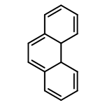
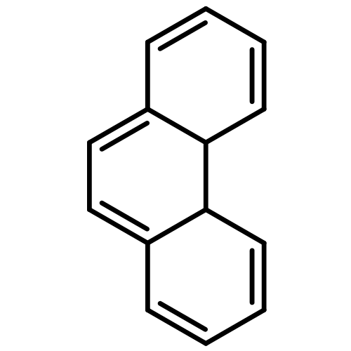
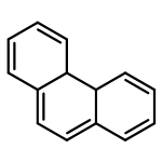
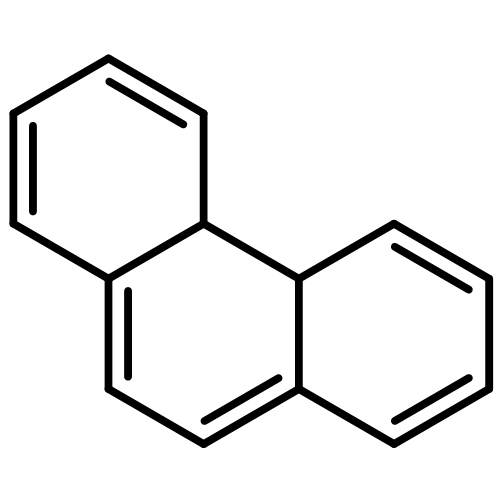
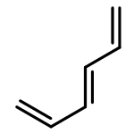
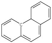
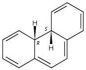
Ultrafast time-resolved mass spectrometry and structural dynamics experiments on trans-stilbene, cis-stilbene, and azobenzene, with excitation to high-lying electronic states, reveal a rich diversity of photochemical reaction dynamics. All processes are found to be quite unlike the well-known photochemistry on lower electronic surfaces. While in trans-stilbene, excitation at 6 eV induces a phenyl twisting motion, in cis-stilbene it leads to an ultrafast ring-closing to form 4a,4b-dihydrophenanthrene. Azobenzene dissociates on an ultrafast time scale, rather than isomerizing as it does on a lower surface. The photochemical dynamics of the sample molecules proceed along steep potential energy surfaces and conical intersections. Because of that, the dynamics are much faster than vibrational relaxation, the randomizing effects from vibrational energy scrambling are avoided, and excitation-energy specific reaction dynamics results.
Co-reporter:Jie Bao, Michael P. Minitti, and Peter M. Weber
The Journal of Physical Chemistry A 2011 Volume 115(Issue 9) pp:1508-1515
Publication Date(Web):February 10, 2011
DOI:10.1021/jp1095322
The ultrafast dynamics of highly excited cis-stilbene (CS) in a molecular beam is explored using femtosecond time-resolved mass spectrometry and structure-sensitive photoelectron spectroscopy. cis-Stilbene is initially pumped by a 6 eV photon to the 71B state and the reaction is followed by ionization with a time-delayed 3 eV probe pulse. Upon excitation, cis-stilbene rapidly decays to the 31B state, where it undergoes a ring-closing reaction to form 4a,4b-dihydrophenanthrene (DHP). Whereas 14% of the ionized CS molecules dissociate one hydrogen atom to form hydrophenanthrene, the ionized DHP molecules completely dehydrogenate in the ion state to produce hydrophenanthrene and phenanthrene with a 1:1 ratio. We determined the lifetimes of the 71B state and the 31B state of CS to be 167 and 395 fs, respectively.
Co-reporter:Sanghamitra Deb, Brian A. Bayes, Michael P. Minitti, and Peter M. Weber
The Journal of Physical Chemistry A 2011 Volume 115(Issue 10) pp:1804-1809
Publication Date(Web):February 22, 2011
DOI:10.1021/jp110905h
Rotations about its three carbon−nitrogen bonds give triethylamine a complex, 3-dimensional potential energy landscape of conformeric structures. Electronic excitation to Rydberg states prepares the molecule in a high-energy, nonequilibrium distribution of such conformers, initiating ultrafast transitions between them. Time-resolved Rydberg electron binding energy spectra, observed using photoionization-photoelectron spectroscopy with ultrashort laser pulses, reveal these time-evolving structures. The time-dependent structural fingerprint spectra are assigned with the aid of a computational analysis of the potential energy landscape. Upon 209 nm electronic excitation to the 3p Rydberg state, triethylamine decays to 3s with a 200 fs time constant. The initially prepared conformer reacts to a mixture of structures with a time constant of 232 fs and settles into a final geometry distribution on a further subpicosecond time scale. The binding energy of the Rydberg electron is found to be an important determinant of the conformeric energy landscape.
Co-reporter:Xiao Liang, Michael G. Levy, Sanghamitra Deb, Joseph D. Geiser, Richard M. Stratt, Peter M. Weber
Journal of Molecular Structure 2010 Volume 978(1–3) pp:250-256
Publication Date(Web):20 August 2010
DOI:10.1016/j.molstruc.2010.02.041
Rydberg Fingerprint Spectroscopy (RFS) has proven to be a powerful method to probe molecular structures, especially transient structures with large amounts of internal energy. The technique is related to electron diffraction in that the molecular structure sensitivity originates from the wavefunction phase shift of the probe electron induced by the charge distributions in the molecule. Exploiting the close relationship between the two techniques, we investigate the origin of the molecular structure sensitivity of RFS by introducing a molecular ion core model that is solved analytically using perturbation theory as well as numerically using a numerical-grid method. The dependence of the Rydberg electron energy on some molecular parameters is investigated and the effectiveness of the numerical-grid method in solving our one-electron Schrödinger equation is validated. A reasonable consistency between experimental and computed quantum defect values is shown for specific molecular ion core model parameters.
Co-reporter:Joseph C. Bush, Michael P. Minitti, and Peter M. Weber
The Journal of Physical Chemistry A 2010 Volume 114(Issue 42) pp:11078-11084
Publication Date(Web):July 1, 2010
DOI:10.1021/jp101881x
Time-resolved multiphoton ionization mass spectrometry coupled with Rydberg Fingerprint Spectroscopy (RFS) has been used to analyze the structural and electronic dynamics of N,N-dimethylphenethylamine (PENNA) and N,N-dimethylcyclohexethylamine (CENNA). In PENNA, the molecule converts from 3p to 3s on a time scale of 149 fs, a process that is reflected in the mass spectrum as the onset of fragmentation. Once in 3s, the overall signal intensity of the PENNA 3s signal shows biexponential decay kinetics, which is attributed to the electronic curve crossing from the Rydberg state to a dissociative antibonding orbital of the ethylenic bridge. This curve crossing exemplifies a possible fragmentation pathway observed in electron capture dissociation of proteins. The initially fast reaction (1.3 ps) is greatly slowed down as a result of an apparent relaxation process with a 5.6 ps time constant. The electron binding energy of the 3s Rydberg state of PENNA is observed to shift with a time constant of 4.8 ps, which is correlated to a cation−π interaction driven conformeric rearrangement.
Co-reporter:Fedor Rudakov and Peter M. Weber
The Journal of Physical Chemistry A 2010 Volume 114(Issue 13) pp:4501-4506
Publication Date(Web):March 12, 2010
DOI:10.1021/jp910786s
We explored the curve crossing dynamics of 1,2,3,4-tetramethyl-cyclopentadiene (TMCPD) and 1,2,3,4,5-pentamethyl-cyclopentadiene (PMCPD) upon π → π* excitation to the 1B2 state using time-resolved, resonance-enhanced multiphoton ionization mass and photoelectron spectroscopy. Upon excitation with a femtosecond laser pulse at 267 nm, the energy relaxation pathway is observed by a time-delayed probe pulse at 400 nm, which ionizes the molecule through Rydberg states that reveal the momentary state of the molecule in the photoelectron spectra. We observe that the initially populated 1B2 state decays to the 2A1 surface in 135 fs in TMCPD and 183 fs in PMCPD, followed by a crossing to the ground state 1A1 surface on 57 and 60 fs time scales for TMCPD and PMCPD, respectively. The spectroscopic signatures of the 2A1 states are clearly revealed in the two-photon ionization photoelectron spectra. In both systems we observe that the ground states are recovered completely, indicating that no new molecular structures are created on the time scale of the experiment.
Co-reporter:Joseph C. Bush, Michael P. Minitti, Peter M. Weber
Journal of Photochemistry and Photobiology A: Chemistry 2010 Volume 213(Issue 1) pp:70-72
Publication Date(Web):10 June 2010
DOI:10.1016/j.jphotochem.2010.05.001
Ultrafast conformeric dynamics in N,N-dimethylphenethylamine is initiated by photo-excitation to the 3p Rydberg level. On the ion-like potential energy surface of the Rydberg state, the formation of an intramolecular cation–pi bond between the positively charged amine ion core and the aromatic ring is observed in real time using Rydberg fingerprint spectroscopy. The structural dynamics of the cation–pi bond formation proceeds with a 5.3 ps time constant at an internal temperature of 760 K.
Co-reporter:Jie Bao and Peter M. Weber
The Journal of Physical Chemistry Letters 2010 Volume 1(Issue 1) pp:224-227
Publication Date(Web):November 20, 2009
DOI:10.1021/jz900147b
Using femtosecond time-resolved resonant two-photon ionization coupled with photoelectron spectroscopy, we have explored the structural dynamics of a previously uncharted, highly excited state of trans-stilbene. The molecule responds to excitation to the S5 surface by twisting its phenyl groups about the carbon−carbon bonds. The motion of the coherent wave packet is reflected in the time-dependent spectrum, yielding an oscillatory frequency of 6.7 cm−1. This structural oscillation competes with a transition to a lower surface with a time constant of 280 (±30) fs. The dynamical motions are intriguingly different from those that have previously been observed in lower excited states, revealing a structural dynamics that strongly depends on the electronic surface.Keywords (keywords): stilbene; structural dynamics; ultrafast photoelectron spectroscopy;
Co-reporter:Fedor Rudakov, Peter M. Weber
Chemical Physics Letters 2009 470(4–6) pp: 187-190
Publication Date(Web):
DOI:10.1016/j.cplett.2009.01.058
Co-reporter:Job D. Cardoza, Fedor M. Rudakov, Nils Hansen, Peter M. Weber
Journal of Electron Spectroscopy and Related Phenomena 2008 Volume 165(1–3) pp:5-10
Publication Date(Web):September 2008
DOI:10.1016/j.elspec.2008.06.003
Many saturated and unsaturated organic hydrocarbons can assume multiple isomeric forms. At high temperatures, the identification of such isomers is difficult with conventional spectroscopic techniques, posing a challenge to the exploration of important processes such as the combustion of hydrocarbons. A recently developed technology using resonance enhanced multi-photon ionization via Rydberg states shows promise as an analytical technique, because the resultant photoelectron spectra provide well-resolved peaks that are sensitive to the molecular structure even at high temperatures. We tested this new method on isomeric hydrocarbon systems with the chemical formulas C5H8, C6H8, C7H8, and C11H10. Three-photon ionization with femtosecond pulses near 400 nm shows that in all systems the observation of distinct Rydberg features can serve to identify the isomers. In the C5H8 system, the photoelectron spectra of the isomers show Rydberg peaks from different quantum states, making the spectral identification of those isomers especially facile. Thus it appears that photoionization from Rydberg states could be developed into an analytical tool that is capable of distinguishing isomeric hydrocarbons even under adverse conditions such as flames and other combustion phenomena.
Co-reporter:Job D. Cardoza, Fedor M. Rudakov and Peter M. Weber
The Journal of Physical Chemistry A 2008 Volume 112(Issue 43) pp:10736-10743
Publication Date(Web):October 4, 2008
DOI:10.1021/jp8041236
Resonance-enhanced multiphoton ionization photoelectron spectroscopy has been applied to study the electronic spectroscopy and relaxation pathways among the 3p and 3s Rydberg states of trimethylamine. The experiments used femtosecond and picosecond duration laser pulses at wavelengths of 416, 266, and 208 nm and employed two-photon and three-photon ionization schemes. The binding energy of the 3s Rydberg state was found to be 3.087 ± 0.005 eV. The degenerate 3px,y states have binding energies of 2.251 ± 0.005 eV, and 3pz is at 2.204 ± 0.005 eV. Using picosecond and femtosecond time-resolved experiments we spectrally and temporally resolved an intricate sequence of energy relaxation pathways leading from the 3p states to the 3s state. With excitation at 5.96 eV, trimethylamine is found to decay from the 3pz state to 3px,y in 539 fs. The decay to 3s from all the 3p states takes place with a 2.9 ps time constant. On these time scales, trimethylamine does not fragment at the given internal energies, which range from 0.42 to 1.54 eV depending on the excitation wavelength and electronic state.
Co-reporter:Job D. Cardoza, Raymond C. Dudek, Richard J. Mawhorter, Peter M. Weber
Chemical Physics 2004 Volume 299(2–3) pp:307-312
Publication Date(Web):19 April 2004
DOI:10.1016/j.chemphys.2003.12.018
Abstract
Pump–probe electron diffraction is an exciting new technique to probe the structural dynamics of gas-phase molecules in transient states. A difficult aspect of this experiment is that pump–probe electron diffraction patterns are sensitive toward minute pointing instabilities of the electron beam. Displacements of the beam in the range of microradians, depending on the electron beam energy, can cause artifacts in the pump–probe difference patterns that are larger than the desired pump–probe diffraction signature. We describe a solution to the problem, which uses a novel centering algorithm that takes advantage of the inherent symmetry of the patterns and that acts only on the molecular component of the diffraction signal. The implementation of the algorithm is illustrated using the pump–probe diffraction signature obtained in the ultrafast time-resolved study of the ring-opening reaction of 1,3-cyclohexadiene to form 1,3,5-hexatriene.
Co-reporter:Sergey A. Shlykov, Tran D. Phien, Yan Gao, Peter M. Weber
Journal of Molecular Structure (15 March 2017) Volume 1132() pp:
Publication Date(Web):15 March 2017
DOI:10.1016/j.molstruc.2016.06.048
•N-phenylpiperidine was studied by GED and quantum chemistry.•In the gas phase the compound exist in equatorial and axial forms.•The equatorial form is dominating, the ΔG° value is 0.7–1.4 kcal/mol.•Orientation of the phenyl group is governed by hyperconjugation.Molecular structure and conformational behavior of N-phenylpiperidine (NPhP) were investigated by synchronous gas-phase electron diffraction/mass spectrometry (GED/MS) and quantum chemistry. Due to influence of steric repulsion and hyperconjugation, NPhP may exist in two conformers, equatorial and axial chair forms. Both experiment and theoretical calculations suggest a C1 symmetry of the conformers, with the plane perpendicular to the phenyl group turned by ca. 30–40° (equatorial) and 0–20° (axial) about the plane perpendicular to the piperidine ring symmetry plane. According to the QC calculations, NPhP may exist as two conformers, equatorial and axial, with a ratio of Eq:Ax = 92:8 (B3LYP), 87:13 (B3LYP-GD3), 84:16 (M06-2X), 83:17 (MP2/6-311G**) and 76:24% (MP2/cc-pVTZ). Except for the latter, these values are in good agreement with the experimental GED data of 90(10):10(10)%.A comparative analysis of similar compounds, phenylcyclohexane and 1-phenylheterocyclohexanes, was performed. Conformational properties depend on the CPhX bond distance and hyperconjugation between the phenyl ring and the lone pair on the heteroatom. The contribution of the axial form of 1-phenylcyclohexane derivatives increases in the series of the heteroatom X in the cyclohexane ring: C → N → Si → P.
Co-reporter:Sergey A. Shlykov, Tran Dinh Phien, Peter M. Weber
Journal of Molecular Structure (15 June 2017) Volume 1138() pp:
Publication Date(Web):15 June 2017
DOI:10.1016/j.molstruc.2017.03.006
•N-cyanopiperidine was studied by GED, IR spectroscopy and quantum chemistry.•The ratio of equatorial and axial forms is 52(6):48(6)% (GED) and 60(6):40(6)% (IR).•The M062X and MP2 calculations are in good agreement with experimental data.•The CCSD(T)/CBS calculations find a value of Eax–Eeq = 0.26 kcal/mol.•Conjugation stabilizes the axial forms of 1-cyano-heterocyclohexanes.The molecular structure and intramolecular inversions of N-cyanopiperidine (CNP) were studied by various quantum chemical methods and using synchronous gas electron diffraction/mass spectrometry (GED/MS) and IR spectroscopy. The contribution of the equatorial conformer was found GED 52(6), IR 60(6), B3LYP 77, B3LYP-D3 67, M062X 59, MP2 54%. Interpolation using CCSD(T)-F12/CBS predicts Eax–Eeq = 0.26 kcal/mol. The NBO analysis shows a strong conjugation of the nitrogen atom lone pair with the triple bond making the r(N–CCN) = 1.355(3) Å distance as short as a typical sesquilateral bond.A low energy barrier for the Eq→Ax transition of 1.0–2.3 kcal/mol in CNP leads to difficulties in NMR conformational ratio studies. This leaves the GED method almost as the only experimental method to study both structural and conformational properties.Conformational properties of the related compounds are found to depend on the X–CCN bond distance and conjugation between the CN group and the lone pair on the heteroatom. The contribution of the axial form of 1-cyano-heterocyclohexanes increases in the series of heteroatoms X in the cyclohexane ring: N→C→Si→P.
Co-reporter:Xinxin Cheng, Yan Gao, Fedor Rudakov and Peter M. Weber
Chemical Science (2010-Present) 2016 - vol. 7(Issue 1) pp:NaN627-627
Publication Date(Web):2015/10/19
DOI:10.1039/C5SC03042K
Three ionization centers make 1,3,5-trimethyl-1,3,5-triazacyclohexane (TMTAC) an interesting model system to study intramolecular charge transfer (CT). Because the molecule assumes a Cs symmetric, axial–equatorial–equatorial (aee) conformation in the ground state, there are two distinct types of the nitrogen atoms. We discovered that either nitrogen atom can be ionized independently so that two molecular cations exist with different (localized) charge distributions in the Franck–Condon region. The initially localized charge can delocalize via CT, provided the molecule acquires a suitable structural geometry. These proper structures are all found to have a common structural motif that supports CT via through-space-interaction. The structural dynamics and the CT process in Rydberg-excited TMTAC, where the molecular ion core closely resembles the ion, were probed by time-resolved Rydberg fingerprint spectroscopy. When TMTAC is excited at 230 nm to the Franck–Condon region of the 3s Rydberg state, the two types of nitrogen atom Rydberg chromophores give rise to distinct binding energy peaks. The sequential molecular responses that follow the Rydberg excitation manifest themselves as time-dependent changes of the binding energy and are observed by ionization at 404 nm. A fast transition with 103 fs time constant was attributed to a motion that leads to a local minimum of the charge-localized state on the Rydberg potential energy surface. Because a large amount of energy is deposited into the vibrational manifolds, the molecule continues to sample the potential energy surface and eventually reaches a dynamic equilibrium between charge-localized and charge-delocalized states. The forward and backward time constants were determined to be 1.02 ps and 4.09 ps, respectively. The binding energy spectrum also reveals the existence of an equilibrium among several charge-delocalized states. Quantum chemical calculations were carried out to find the stable minima of the ground state and the ion state. The binding energies of the Franck–Condon structures and the relaxed ion structures were calculated using the Perdew–Zunger self-interaction corrected DFT (PZ-SIC) method to assign the spectra at time zero and at equilibrium, respectively.
Co-reporter:Xinxin Cheng, Yao Zhang, Sanghamitra Deb, Michael P. Minitti, Yan Gao, Hannes Jónsson and Peter M. Weber
Chemical Science (2010-Present) 2014 - vol. 5(Issue 11) pp:NaN4403-4403
Publication Date(Web):2014/07/16
DOI:10.1039/C4SC01646G
Two nitrogen atoms and a flexible carbon skeleton make N,N,N′,N′-tetramethylethylenediamine (TMEDA) an important model system to study the interplay of conformeric motions and charge delocalization. Ionization of one of the nitrogen atoms generates a localized charge that may (partially) transfer to the other nitrogen. The structural motions, conformation dependent electron lone pair interaction and charge transfer in Rydberg-excited TMEDA, where the molecular ion core closely resembles the ion, were probed by time-resolved Rydberg fingerprint spectroscopy. Excitation to the 3p Rydberg level with a 209 nm laser pulse initially created a charge-localized ion core. Rapid internal conversion to the 3s Rydberg state yielded a multitude of conformational structures, in particular structures that are close to the folded GG′G+ and GGG′+ (see text for label definitions) core structures (235 fs), and structures that are close to the extended TTT+ core structure (557 fs). The initial excitation and the internal conversion deposit about 1.89 eV of energy into the vibrational manifold, enabling a fast equilibrium between the folded and the extended structures. The forward and backward time constants were determined to be 490 fs and 621 fs, respectively. With the molecule highly vibrationally excited, the decay to 3s proceeds with a 6.77 ps time constant. Density functional theory (DFT) and ab initio calculations show evidence of strong lone pair interaction and charge delocalization in the equilibrium conformers. Importantly, DFT with self-interaction correction properly describes the binding energy of the Rydberg electron and provides excellent agreement with the experimental results.


![Phenyl, 2-[(1Z)-2-phenylethenyl]-](http://img.cochemist.com/ccimg/335400/335305-94-7.png)
![Phenyl, 2-[(1Z)-2-phenylethenyl]-](http://img.cochemist.com/ccimg/335400/335305-94-7_b.png)