Co-reporter:Xiaolong Qiu;Trisha M. Westerhof;Amrith A. Karunaratne;Erik M. Werner;Pedram P. Pourfard;Edward L. Nelson;Elliot E. Hui;Jered B. Haun
Lab on a Chip (2001-Present) 2017 vol. 17(Issue 19) pp:3300-3309
Publication Date(Web):2017/09/26
DOI:10.1039/C7LC00575J
The ability to harvest single cells from tissues is currently a bottleneck for cell-based diagnostic technologies, and remains crucial in the fields of tissue engineering and regenerative medicine. Tissues are typically broken down using proteolytic digestion and various mechanical treatments, but success has been limited due to long processing times, low yield, and high manual labor burden. Here, we present a novel microfluidic device that utilizes precision fluid flows to improve the speed and efficiency of tissue digestion. The microfluidic channels were designed to apply hydrodynamic shear forces at discrete locations on tissue specimens up to 1 cm in length and 1 mm in diameter, thereby accelerating digestion through hydrodynamic shear forces and improved enzyme–tissue contact. We show using animal organs that our digestion device with hydro-mincing capabilities was superior to conventional scalpel mincing and digestion based on recovery of DNA and viable single cells. Thus, our microfluidic digestion device can eliminate or reduce the need to mince tissue samples with a scalpel, while reducing sample processing time and preserving cell viability. Another advantage is that downstream microfluidic operations could be integrated to enable advanced cell processing and analysis capabilities. We envision our novel device being used in research and clinical settings to promote single cell-based analysis technologies, as well as to isolate primary, progenitor, and stem cells for use in the fields of tissue engineering and regenerative medicine.
Co-reporter:Mingqiu Wang, Shreyas R. Ravindranath, Maha K. Rahim, Elliot L Botvinick, and Jered B. Haun
Langmuir 2016 Volume 32(Issue 49) pp:13124-13136
Publication Date(Web):October 31, 2016
DOI:10.1021/acs.langmuir.6b03014
The targeted delivery of nanoparticle carriers holds tremendous potential to transform the detection and treatment of diseases. A major attribute of nanoparticles is the ability to form multiple bonds with target cells, which greatly improves the adhesion strength. However, the multivalent binding of nanoparticles is still poorly understood, particularly from a dynamic perspective. In previous experimental work, we studied the kinetics of nanoparticle adhesion and found that the rate of detachment decreased over time. Here, we have applied the adhesive dynamics simulation framework to investigate binding dynamics between an antibody-conjugated, 200-nm-diameter sphere and an ICAM-1-coated surface on the scale of individual bonds. We found that nano adhesive dynamics (NAD) simulations could replicate the time-varying nanoparticle detachment behavior that we observed in experiments. As expected, this behavior correlated with a steady increase in mean bond number with time, but this was attributed to bond accumulation only during the first second that nanoparticles were bound. Longer-term increases in bond number instead were manifested from nanoparticle detachment serving as a selection mechanism to eliminate nanoparticles that had randomly been confined to lower bond valencies. Thus, time-dependent nanoparticle detachment reflects an evolution of the remaining nanoparticle population toward higher overall bond valency. We also found that NAD simulations precisely matched experiments whenever mechanical force loads on bonds were high enough to directly induce rupture. These mechanical forces were in excess of 300 pN and primarily arose from the Brownian motion of the nanoparticle, but we also identified a valency-dependent contribution from bonds pulling on each other. In summary, we have achieved excellent kinetic consistency between NAD simulations and experiments, which has revealed new insights into the dynamics and biophysics of multivalent nanoparticle adhesion. In future work, we will leverage the simulation as a design tool for optimizing targeted nanoparticle agents.
Co-reporter:Xiaolong Qiu, Janice De Jesus, Marissa Pennell, Marco Troiani and Jered B. Haun
Lab on a Chip 2015 vol. 15(Issue 1) pp:339-350
Publication Date(Web):29 Oct 2014
DOI:10.1039/C4LC01126K
Tumors tissues house a diverse array of cell types, requiring powerful cell-based analysis methods to characterize cellular heterogeneity and identify rare cells. Tumor tissue is dissociated into single cells by treatment with proteolytic enzymes, followed by mechanical disruption using vortexing or pipetting. These procedures can be incomplete and require significant time, and the latter mechanical treatments are poorly defined and controlled. Here, we present a novel microfluidic device to improve mechanical dissociation of digested tissue and cell aggregates into single cells. The device design includes a network of branching channels that range in size from millimeters down to hundreds of microns. The channels also contain flow constrictions that generate well-defined regions of high shear force, which we refer to as “hydrodynamic micro-scalpels”, to progressively disaggregate tissue fragments and clusters into single cells. We show using in vitro cancer cell models that the microfluidic device significantly enhances cell recovery in comparison to mechanical disruption by pipetting and vortexing after digestion with trypsin or incubation with EDTA. Notably, the device enabled superior results to be obtained after shorter proteolytic digestion times, resulting in fully viable cells in less than ten minutes. The device could also be operated under enzyme-free conditions that could better maintain expression of certain surface markers. The microfluidic format is advantageous because it enables application of well-defined mechanical forces and rapid processing times. Furthermore, it may be possible to directly integrate downstream processing and detection operations to create integrated cell-based analysis platforms. The enhanced capabilities enabled by our novel device may help promote applications of single cell detection and purification techniques to tumor tissue specimens, advancing the current understanding of cancer biology and enabling molecular diagnostics in clinical settings.
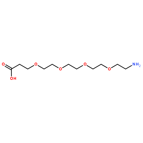
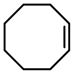
Co-reporter:Maha K. Rahim, Rajesh Kota, and Jered B. Haun
Bioconjugate Chemistry 2015 Volume 26(Issue 2) pp:352
Publication Date(Web):January 13, 2015
DOI:10.1021/bc500605g
The bioorthogonal cycloaddition reaction between tetrazine and trans-cyclooctene (TCO) is rapidly growing in use for molecular imaging and cell-based diagnostics. We have surprisingly uncovered that the majority of TCOs conjugated to monoclonal antibodies using standard amine-coupling procedures are nonreactive. We show that antibody-bound TCOs are not inactivated by trans–cis isomerization and that the bulky cycloaddition reaction is not sterically hindered. Instead, TCOs are likely masked by hydrophobic interactions with the antibody. We show that introducing TCO via hydrophilic poly(ethylene glycol) (PEG) linkers can fully preserve reactivity, resulting in >5-fold enhancement in functional density without affecting antibody binding. This is accomplished using a novel dual bioorthogonal approach in which heterobifunctional dibenzylcyclooctyne (DBCO)–PEG–TCO molecules are reacted with azido-antibodies. Improved imaging capabilities are demonstrated for different cancer biomarkers using tetrazine-modified fluorophore and quantum dot probes. We believe that the PEG linkers prevent TCOs from burying within the antibody during conjugation, which could be relevant to other bioorthogonal tags and biomolecules. We expect the improved TCO reactivity obtained using the reported methods will significantly advance bioorthogonal pretargeting applications.
Co-reporter:Jered B. Haun, Lauren R. Pepper, Eric T. Boder, and Daniel A. Hammer
Langmuir 2011 Volume 27(Issue 22) pp:13701-13712
Publication Date(Web):September 26, 2011
DOI:10.1021/la202926m
Elucidation of the relationship between targeting molecule binding properties and the adhesive behavior of therapeutic or diagnostic nanocarriers would aid in the design of optimized vectors and lead to improved efficacy. We measured the adhesion of 200-nm-diameter particles under fluid flow that was mediated by a diverse array of molecular interactions, including recombinant single-chain antibodies (scFvs), full antibodies, and the avidin/biotin interaction. Within the panel of scFvs, we used a family of mutants that display a spectrum of binding kinetics, allowing us to compare nanoparticle adhesion to bond chemistry. In addition, we explored the effect of molecular size by inserting a protein linker into the scFv fusion construct and by employing scFvs that are specific for targets with vastly different sizes. Using computational models, we extracted multivalent kinetic rate constants for particle attachment and detachment from the adhesion data and correlated the results to molecular binding properties. Our results indicate that the factors that increase encounter probability, such as adhesion molecule valency and size, directly enhance the rate of nanoparticle attachment. Bond kinetics had no influence on scFv-mediated nanoparticle attachment within the kinetic range tested, however, but did appear to affect antibody/antigen and avidin/biotin mediated adhesion. We attribute this finding to a combination of multivalent binding and differences in bond mechanical strength between recombinant scFvs and the other adhesion molecules. Nanoparticle detachment probability correlated directly with adhesion molecule valency and size, as well as the logarithm of the affinity for all molecules tested. On the basis of this work, scFvs can serve as viable targeting receptors for nanoparticles, but improvements to their bond mechanical strength would likely be required to fully exploit their tunable kinetic properties and maximize the adhesion efficiency of nanoparticles that bear them.
.jpg)