Co-reporter:Lingmei Li, Chengjian Wang, Shan Qiang, Jixiang Zhao, Shuang Song, Wanjun Jin, Bo Wang, Ying Zhang, Linjuan Huang, and Zhongfu Wang
Journal of Agricultural and Food Chemistry 2016 Volume 64(Issue 39) pp:7367-7376
Publication Date(Web):September 12, 2016
DOI:10.1021/acs.jafc.6b02773
Glycosylation of many proteins has been revealed to be closely related with food allergies, and screening and structural analysis of related glycoproteins and glycoallergens are essential for studies in this field. Herein, we describe detailed N-glycoform analysis of all glycoprotein fractions of soybean protein isolate (SPI) separated by sodium dodecyl sulfate polyacrylamide gel electrophoresis (SDS-PAGE) to disclose structural features of the glycan moieties of more soybean glycoproteins. SPI was fractionated by SDS-PAGE, and the generated protein bands were recovered and subjected to in-gel N-glycan release and labeling using a one-pot method newly developed by our group, followed by detailed analysis by electrospray ionization mass spectrometry (ESI-MS) and online hydrophilic interaction liquid chromatography coupled with electrospray ionization tandem mass spectrometry (HILIC-ESI-MS/MS). As a result, we found seven bands mainly containing oligomannose-type glycans; two mainly contain core α1,3-fucosylated glycans, and six have no glycans. This study is the first report that discovers core α1,3-fucosylated N-glycans in bands 1, 2, and 6 and discloses bands 3, 4, 5, and 7 as glycoproteins and their N-glycoforms. Therefore, it can expand our knowledge about soybean protein glycosylation and provide significant structural reference for research of soybean allergens.Keywords: ESI-MS; glycoprotein; HILIC-ESI-MS/MS; N-glycans; soybean allergy;
Co-reporter:Yujiao Sun, Bingying Yang, Yanmin Wu, Yang Liu, Xiao Gu, Hong Zhang, Chengjian Wang, Hongzhi Cao, Linjuan Huang, Zhongfu Wang
Food Chemistry 2015 Volume 178() pp:311-318
Publication Date(Web):1 July 2015
DOI:10.1016/j.foodchem.2015.01.105
•κ-Carrageenan was digested by four different degradation methods.•ESI-MS spectra were used to identify structure and composition of the hydrolysates.•The structures of κ-carrageenan hydrolysates were variant and different.•Antioxidant activities of the κ-carrageenan hydrolysates were evaluated.•H2O2 hydrolysates showed much stronger antioxidant abilities.In the present study, four kinds of κ-carrageenan oligosaccharides were obtained by the degradation of parent κ-carrageenan using free radical depolymerization, mild acid hydrolysis, κ-carrageenase digestion and partial reductive hydrolysis, respectively. Their structure types were accurately and comparatively elucidated by ESI-MS and CID MS/MS. The antioxidant activities of different degraded products were investigated by four different antioxidant assays, including superoxide radical scavenging activity, hydroxyl radical scavenging activity, reducing power and DPPH radical scavenging activity. The methods of depolymerization had an influence on the antioxidant activities of κ-carrageenan oligosaccharides obtained from κ-carrageenan. These results indicated that the antioxidant activities of κ-carrageenan oligosaccharides could be related to the degree of polymerization, the content of reducing sugar and sulfate groups, and the structure of reducing terminus.
Co-reporter:Yujiao Sun, Wei Sun, Jiatong Guo, Xiuting Hu, Guiping Gong, Linjuan Huang, Hongzhi Cao, Zhongfu Wang
Food Chemistry 2015 170() pp: 22-29
Publication Date(Web):
DOI:10.1016/j.foodchem.2014.08.024
Co-reporter:Chengjian Wang, Zhiyu Wu, Jiangbei Yuan, Bo Wang, Ping Zhang, Ying Zhang, Zhongfu Wang, and Linjuan Huang
Journal of Proteome Research 2014 Volume 13(Issue 2) pp:372-384
Publication Date(Web):2017-2-22
DOI:10.1021/pr4010647
Fast, sensitive, and simple methods for quantitative analysis of disparities in glycan expression between different biological samples are essential for studies of protein glycosylation patterns (glycomics) and the search for disease glycan biomarkers. Relative quantitation of glycans based on stable isotope labeling combined with mass spectrometric detection represents an emerging and promising technique. However, this technique is undermined by the complexity of mass spectra of isotope-labeled glycans caused by the presence of multiple metal ion adduct signals, which result in a decrease of detection sensitivity and an increase of difficulties in data interpretation. Herein we report a simplified quantitative glycomics strategy, which features nonreductive isotopic labeling of reducing glycans with either nondeuterated (d0-) or deuterated (d5-) Girard’s reagent P (GP) without salts introduced and simplified mass spectrometric profiles of d0- and d5-GP derivatives of neutral glycans as molecular ions without complex metal ion adducts, allowing rapid and sensitive quantitative comparison between different glycan samples. We have obtained optimized GP-labeling conditions and good quantitation linearity, reproducibility, and accuracy of data by the method. Its excellent applicability was validated by comparatively quantitative analysis of the neutral N-glycans released from bovine and porcine immunoglobulin G as well as of those from mouse and rat sera. Additionally, we have revealed the potential of this strategy for the high-sensitivity analysis of sialylated glycans as GP derivatives, which involves neutralization of the carboxyl group of sialic acid by chemical derivatization.
Co-reporter:Kuan Jiang, Chengjian Wang, Yujiao Sun, Yang Liu, Ying Zhang, Linjuan Huang, and Zhongfu Wang
Journal of Agricultural and Food Chemistry 2014 Volume 62(Issue 29) pp:7245-7254
Publication Date(Web):July 7, 2014
DOI:10.1021/jf501352j
Species-specific ovotransferrin features a highly conservative protein sequence, but it varies in the structure of the attached oligosaccharides, which may contribute to the differences observed in its bioactivity and nutritional value. Herein, chicken ovotransferrin (COT) and pheasant ovotransferrin (POT) isolated by repeated ethanol precipitation of egg white were digested with peptide N-glycosidase F to release N-glycans. The obtained N-glyans were isotopically labeled with aniline and analyzed via electrospray ionization mass spectrometry and online hydrophilic interaction liquid chromatography coupled with tandem mass spectrometry (HILIC–MS/MS). Relative quantitation based on isotopic aniline labeling and HILIC–MS/MS analysis revealed in detail the conspicuous difference between COT and POT in the abundance of their N-glycan compositions and isomers. In total, 16 COT N-glycans were observed, including 1 core structure (3.18%), 3 hybrid type (5.42%), and 12 complex type (91.40%), whereas 21 POT N-glycans were found, including 1 truncated structure (1.88%), 1 core structure (6.26%), 3 high mannose type (5.20%), 6 hybrid type (19.14%), and 10 complex type (67.52%). To our knowledge, this study is the first qualitative and quantitative comparison of COT and POT N-glycosylation patterns. These results suggest that POT has a different glycosylation pattern compared to that of COT and thus the effect of its glycosylation pattern on its bioactivity is worthy of further exploration.
Co-reporter:Jiangbei Yuan, Chengjian Wang, Yujiao Sun, Linjuan Huang, Zhongfu Wang
Analytical Biochemistry 2014 Volume 462() pp:1-9
Publication Date(Web):1 October 2014
DOI:10.1016/j.ab.2014.05.029
Abstract
A novel strategy is proposed, using cost-saving chemical reactions to generate intact free reducing N-glycans and their fluorescent derivatives from glycoproteins for subsequent analysis. N-Glycans without core α-1,3-linked fucose are released in reducing form by selective hydrolysis of the N-type carbohydrate–peptide bond of glycoproteins under a set of optimized mild alkaline conditions and are comparable to those released by commonly used peptide-N-glycosidase (PNGase) F in terms of yield without any detectable side reaction (peeling or deacetylation). The obtained reducing glycans can be routinely derivatized with 2-aminobenzoic acid (2-AA), 1-phenyl-3-methyl-5-pyrazolone (PMP), and potentially some other fluorescent reagents for comprehensive analysis. Alternatively, the core α-1,3-fucosylated N-glycans are released in mild alkaline medium and derivatized with PMP in situ, and their yields are comparable to those obtained using commonly used PNGase A without conspicuous peeling reaction or any detectable deacetylation. Using this new technique, the N-glycans of a series of purified glycoproteins and complex biological samples were successfully released and analyzed by electrospray ionization mass spectrometry (ESI–MS) and tandem mass spectrometry (MS/MS), demonstrating its general applicability to glycomic studies.
Co-reporter:Ying Zhang;Chengjian Wang;Yang Liu;Weinan Yao
European Food Research and Technology 2014 Volume 239( Issue 5) pp:867-875
Publication Date(Web):2014 November
DOI:10.1007/s00217-014-2283-z
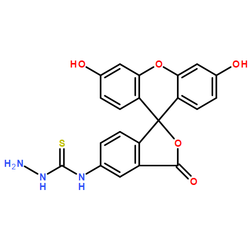
Confocal microscopy is a powerful and effective tool for in vivo elucidation of the molecular basis of biological molecules. Chemical labeling of non-ultraviolet or non-fluorescent carbohydrate with fluorescent tag is an essential step that makes intra-cellular microscopic inspection possible. However, current labeling methods have proved unsuitable for functional pectin-derived oligogalacturonic acid due to the low fluorescence intensity and/or the formation of lactone byproducts. Here, fluorescein-5-thiosemicarbazide (FTSC) was employed for fluorescent labeling of pectin-derived oligogalacturonic acid under mild and neutral reaction conditions to avoid the generation of lactone byproducts. ESI–MS and LC–MS data displayed that pectin-derived pentagalacturonic acid, GalA5, could be quantitatively labeled with FTSC to afford thiosemicarbazones, without any lactone byproducts yielded. Moreover, GalA5-FTSC thiosemicarbazone showed good stability and desirable fluorescence characteristics and exhibited rather low levels of cytotoxicity against sensitive rat thymus cells. Additionally, we have achieved successful fluorescent imaging of GalA5-FTSC thiosemicarbazones transported in living SW-620 and HepG2 cells. The assay illustrates methodological significance in structure–activity elucidation study of functional carbohydrates especially acid oligo-/polysaccharides.
Co-reporter:Chengjian Wang, Jiangbei Yuan, Zhongfu Wang, Linjuan Huang
Journal of Chromatography A 2013 Volume 1274() pp:107-117
Publication Date(Web):25 January 2013
DOI:10.1016/j.chroma.2012.12.005
Qualitative and quantitative analysis of naturally-occurring complex glycans is essential for glycomics, which focuses on the studies of structure–function correlations of saccharides. We previously reported a one-pot procedure for the non-reductive release from glycoproteins and in situ labeling with 1-phenyl-3-methyl-5-pyrazolone (PMP) of O-glycans (C. Wang et al., Proteomics 11 (2011) 4229). Here we describe an HPLC-based O-glycan analytical strategy that combines a range of techniques including the one-pot procedure, independent separation by hydrophilic interaction liquid chromatography (HILIC) and reversed-phase high-performance liquid chromatography (RP-HPLC) and identification by electrospray ionization mass spectrometry (ESI-MS) and tandem mass spectrometry (MS/MS). The complex mixtures of both neutral and sialylated O-glycans as bis-PMP derivatives released from glycoproteins using the one-pot procedure could be well separated by HILIC based on their size or by RP-HPLC depending on the type of linkage and their resulting three-dimensional (3D) structure, and their structure could be characterized by the ESI-MS and MS/MS analysis of the eluted glycan fractions. The validity of the current strategy was confirmed by the analysis of O-glycans released by the one-pot procedure from some standard glycoproteins, including porcine stomach mucin (PSM), bovine submaxillary mucin (BSM) and bovine fetuin. The applicability of the current method to complex biological samples was also demonstrated by the analysis of mucin-type glycans from fetal bovine serum (FBS) and frog egg–jelly coat (FEC). This strategy, a powerful analytical tool that features the combination of different techniques, is useful for the qualitative and quantitative O-glycan analysis of more complex biological samples and has a potential for constructing an O-glycan analysis and structure database.Highlights► The current strategy is powerful due to the combination of different techniques. ► PMP derivatives of O-glycans are separated by HILIC depending on their size. ► PMP derivatives of O-glycans are separated by RP-HPLC based on their 3D-structure. ► This strategy has advantages over conventional O-glycan analysis methods. ► This strategy could be utilized for constructing an O-glycan analysis database.
Co-reporter:Chengjian Wang, Jiangbei Yuan, Xiaohua Li, Zhongfu Wang and Linjuan Huang
Analyst 2013 vol. 138(Issue 18) pp:5344-5356
Publication Date(Web):24 Jun 2013
DOI:10.1039/C3AN00931A
Qualitative and quantitative studies of glycosylation patterns of various biologically important proteins represent a key field for the understanding of their complex structure–function relationships. However, the analysis of glycoprotein glycans is usually undermined by tedious sample processing steps prior to detection, including deproteination, desalting and removal of some other non-glycan impurities, which results in considerable sample loss and increased difficulty of quantitative analysis. Herein we report a facile and versatile method for the quantitative isolation of reducing glycans from complex samples using sulfonyl hydrazine-functionalized polystyrene (SHPS) beads, namely the SHPS-based glycan capturing procedure. This method allows the chemoselective and efficient condensation of the aldehyde group of reducing glycans with the active sulfonyl hydrazine group of SHPS beads under anhydrous conditions, resulting in the formation of sulfonyl hydrazone conjugates. The non-glycan components in samples, such as proteins, salts and some other impurities, can be completely removed by washing the sulfonyl hydrazone conjugates. Regeneration of the reducing glycans can be performed via mild hydrolysis of the washed sulfonyl hydrazone conjugates. This procedure is compatible with almost all the current techniques for the derivatization or detection of reducing glycans. We have obtained essential data for this method, including optimized reaction conditions, linearity and reproducibility for glycan quantitation, as well as a final glycan recovery ratio. Moreover, mass spectrometric analysis of the glycans from some complex biological samples, including milk, human blood plasma and fetal bovine serum, was achieved, demonstrating good applicability of this novel procedure.
Co-reporter:Xiaopeng Lv, Chengjian Wang, Yang Cheng, Linjuan Huang, Zhongfu Wang
Carbohydrate Research 2013 Volume 365() pp:20-25
Publication Date(Web):10 January 2013
DOI:10.1016/j.carres.2012.10.013
A complex polysaccharide, termed LRP4-A, was isolated from the fruit of Lycium ruthenicum Murr. and its structure was characterized. The crude polysaccharide LRP was obtained from the fruit of L. ruthenicum Murr. using hot water extraction followed by ethanol precipitation. The water-soluble polysaccharide LRP4-A was purified from LRP by anion-exchange chromatography and gel filtration chromatography. Its molecular weight was 1.05 × 10⁵ Da. Monosaccharide composition analysis revealed that LRP4-A mainly consisted of rhamnose, arabinose, glucose, and galactose in the molar ratio of 1:7.6:0.5:8.6, with a trace of xylose. Structure of the polysaccharide LRP4-A was characterized using a series of analytical techniques, including methylation analysis, partial acid hydrolysis, IR, NMR, and ESI-MS. LRP4-A was identified to be a highly branching polysaccharide with a backbone of β-(1→6)-linked galactose partially substituted at O-3 position. The branches were composed of (1→3)-linked-Gal, (1→3)-linked-Ara, (1→5)-linked-Ara, and (1→2,4)-linked-Rha. Arabinose, galactose, and glucose were located at the termini of the branches.Graphical abstractHighlights► A homogeneous polysaccharide is isolated from Lycium ruthenicum Murr. ► LRP4-A is composed of Rha, Ara, Xyl, Glc, and Gal, and its MW is 1.05 × 10⁵ Da. ► LRP4-A is a highly branching polysaccharide with a backbone of β-(1→6)-linked-Gal. ► The branches are (1→3) Gal, (1→3) Ara, (1→5) Ara, and (1→2,4) Rha.
Co-reporter:Ying Zhang, Zhongfu Wang, Xiaorui Zhang, Wenxia Zhou, Linjuan Huang
Carbohydrate Research 2011 Volume 346(Issue 14) pp:2156-2164
Publication Date(Web):18 October 2011
DOI:10.1016/j.carres.2011.07.014
A simple and efficient procedure for the fluorescent labeling of saccharides is a prerequisite step for imaging the transport of polysaccharides in living cells. We report a one-pot strategy for the fluorescent labeling of saccharides with fluorescein-5-thiosemicarbazide (FTSC), which introduces the thiosemicarbazide group of FTSC to the aldehyde group at the reducing end of saccharides to form stable amino derivatives via reductive amination. The Glc-FTSC derivative was characterized by HPLC–MS, HRESIMS and NMR spectroscopy. Saccharides were quantitatively labeled with FTSC at 75 °C for 1 h under optimum reaction conditions. Fluorescence studies illustrated that the conjugation of FTSC to saccharides did not change its florescence properties (λex = 495 nm, λem = 517 nm), presenting desirable compatibility with commonly used fluorescence equipment. Polysaccharide AAG-FTSC derivatives exhibited rather low levels of cytotoxicity against rat thymus cells, and the fluorescent labeling procedure had slight impact on their anti-tumor activity. Results indicate that the assay neither introduces discernible cytotoxicity against living cells nor obviously alters the functional activities of polysaccharides, and provides a convenient, highly efficient fluorescent labeling approach for imaging the transport of polysaccharides in living cells.
Co-reporter:Ping Zhang, Ying Zhang, Xiangdong Xue, Chenjian Wang, Zhongfu Wang, Linjuan Huang
Analytical Biochemistry 2011 418(1) pp: 1-9
Publication Date(Web):
DOI:10.1016/j.ab.2011.07.006
Co-reporter:Ying ZHANG;Lin-Juan HUANG;Zhong-Fu WANG
Chinese Journal of Chemistry 2007 Volume 25(Issue 10) pp:1522-1528
Publication Date(Web):16 OCT 2007
DOI:10.1002/cjoc.200790280
3-Amino-9-ethylcarbazole (AEC) was employed for monosaccharide derivatization. The derivatives can be analyzed by high-performance liquid chromatography (HPLC) with an ultraviolet detection (wavelength: 254 nm). Monosaccharides were quantitatively derivatized with 3-amino-9-ethylcarbazole (AEC) at 70 °C for 60 min. The method was linear for all samples over the concentration range tested (r>0.999), the precision was found to be satisfactory (R.S.D.<3%), and the recovery ratios were >98.62%. The stability analysis showed R.S.D. was between 1.81% –3.16%. Detection limits for the samples (D-glucose, L-xylose, D-mannose, L-arabinose, and L-rhamnose) ranged from 0.06 to 1.97 ng/mL (S/N=3). Under the optimized derivatization and HPLC conditions, five monosaccharides were well separated using a narrow bore C18 column (250 mm×4.6 mm) with 0.1 mol/L ammonium acetate containing 25% acetonitrile at a flow rate of 0.5 mL/min. As an application, the method has been successfully applied to the determination of monosaccharide compositions of three polysaccharides SPPA-1, SPPB-1 and SPPC-1 of Spirulina platensis. This method also has potential application to oligosaccharide or glycan analyses.
Co-reporter:Wenlong Qiu;Wanli Nie;Yuna Guo;Linjuan Huang
Chromatographia 2007 Volume 66( Issue 11-12) pp:935-939
Publication Date(Web):2007 December
DOI:10.1365/s10337-007-0422-4
Sucralose (1,6-dichloro-1,6-dideoxy-β-d-fructofuranosyl-4-chloro-4-deoxy-α-d-galactopyranoside) is a high-intensity non-nutritive sweetener derived from sucrose. Determination of sucralose in food is important to ensure consistent product quality. The authors have developed a new method for determination of sucralose. The sucralose was converted into its trimethylsilyl (TMS) ether and qualitative and quantitative analysis were achieved by GC–MS and GC–FID, respectively, using myo-inositol ester as the internal standard. A good linear relationship between response and amount of sucralose TMS ether was obtained in the range 0.005–0.06 mg mL−1 (r = 0.9994). The detection limit was 0.25 ng.
Co-reporter:Guiping Gong, Jiangbo Fan, Yujiao Sun, Yanmin Wu, Yang Liu, Wei Sun, Ying Zhang, Zhongfu Wang
Process Biochemistry (February 2016) Volume 51(Issue 2) pp:314-324
Publication Date(Web):1 February 2016
DOI:10.1016/j.procbio.2015.11.013
•A novel polysaccharide (LBLP5-A) was purified from Lycium barbarum leaves.•The structure of LBLP5-A was determined by several chemical methods combined with electrospray ionization–mass spectrometry (ESI–MS).•LBLP5-A exhibited a good scavenging ability on superoxide, hydroxyl, and 1,1-diphenyl-2-picrylhydrazyl (DPPH) radicals.A water-soluble polysaccharide, LBLP5-A, was isolated from Lycium barbarum leaves, which is widely used as a medicine in Asia. The average molecular weight of LBLP5-A is 11.33 × 10⁴ Da. With O-type carbohydrate–peptide linkage in its structure, LBLP5-A afforded glycan fractions of LBLP5-A-OL1, LBLP5-A-OL2, LBLP5-A-OL3, and LBLP5-A-OL4 acquired by β-elimination reaction. Detailed structure patterns of LBLP5-A-OL1 were characterized by monosaccharide composition analysis, methylation analysis, partial acid hydrolysis, Smith degradation, one- or two-dimensional (1D/2D) nuclear magnetic resonance (NMR), and electrospray ionization–mass spectrometry (ESI–MS). LBLP5-A-OL1 was identified to be a highly branched polysaccharide with a backbone of (1 → 3)-linked Gal, which was partially substituted at its O-6 position. These branches, with arabinose and galactose terminals, were assigned to be (1 → 3)-linked Gal, (1 → 4)-linked Gal, (1 → 3)-linked Araf, (1 → 5)-linked Araf, and (1 → 2, 4)-linked Rhaf. LBLP5-A-OL3 and LBLP5-A-OL4 were determined to be a series of oligosaccharides that share the general structure pattern of Aran1 → Galn2 → Rhan3 (n1 = 0–6, n2 = 0–8, n3 = 0–1) by ESI–MS. Moreover, LBLP5-A exhibited excellent antioxidativity, concerning the scavenging abilities on 1,1-diphenyl-2-picrylhydrazyl (DPPH), superoxide, and hydroxyl radicals in vitro .A water-soluble polysaccharide, termed as LBLP5-A, was isolated from Lycium barbarum leaves, which is a well-known medical drug in Asia. Detailed structure patterns and anti-oxidant activity of LBLP5-A-OL1 were characterized.Download full-size image
Co-reporter:Chengjian Wang, Ping Zhang, Wanjun Jin, Lingmei Li, Shan Qiang, Ying Zhang, Linjuan Huang, Zhongfu Wang
Journal of Proteomics (6 January 2017) Volume 150() pp:18-30
Publication Date(Web):6 January 2017
DOI:10.1016/j.jprot.2016.08.012
Co-reporter:Hong Zhang, Hai-Feng Wang, Yang Liu, Lin-Juan Huang, Zhong-Fu Wang, Ye Li
Journal of Ethnopharmacology (3 July 2014) Volume 154(Issue 3) pp:818-824
Publication Date(Web):3 July 2014
DOI:10.1016/j.jep.2014.05.008
Ethnopharmacological relevanceThe rhizome of Panax japonicus C. A. Mey. var. major (Burk.) C. Y. Wu et K. M. Feng (PJ) is a commonly used traditional Chinese medicine to promote hematopoietic effects, promote blood circulation and supporting healthy energy. Aim of the study is to investigate the haematopoietic effects of PJ and determine the mechanism of its haematopoietic activity.Materials and methodsThe crude extract from PJ (PJE) was separated into two fractions: polysaccharides (PJPS) and low-molecular-weight compounds (PJSM). PJPS, and PJSM were incubated with mice spleen cells, and their haematopoietic activities were evaluated by determining the haematopoietic growth factor levels (HGFs) in vitro. The in vivo experiments used anaemia model mice that were given hypodermic injections of N-acetyl phenylhydrazine (APH) and intraperitoneal injections of cyclophosphamide (CTX).ResultsBoth PJPS and PJSM were significantly involved in the haematopoietic effect of PJE. The administration of PJPS and PJSM could accelerate the recovery of the white blood cell (WBC), red blood cell (RBC), and haemoglobin (HGB) levels in the blood deficiency model mice. Haematopoietic activity may result from stimulating the secretion of interleukin-3 (IL-3), interleukin-6 (IL-6), erythropoietin (EPO), GM colony-stimulating factor (CSF), and M-CSF and by the resistance of spleen cells to apoptosis.ConclusionsThe study results support the potential use of PJPS and PJSM for the treatment of anaemia.Download high-res image (157KB)Download full-size image