Co-reporter:Jing Wang, Dai-Xin Ye, Guo-Hai Liang, Jian Chang, Ji-Lie Kong and Ji-Yao Chen
Journal of Materials Chemistry A 2014 vol. 2(Issue 27) pp:4338-4345
Publication Date(Web):02 May 2014
DOI:10.1039/C4TB00366G
We report a novel method for synthesizing water-dispersible silicon nanoparticles (Si NPs) with a simple one-step procedure using mild reagents (3-aminopropyl) trimethoxysilane (APTES) and ascorbate sodium (AS). This is the first report of “green” synthesis of Si NPs on a large scale and at low cost. The method involves a quick reaction in a commonly used round bottom flask at room temperature and pressure without additional treatment and any special equipment. The as-prepared Si NPs have an average diameter of 2 nm and an emission band at 530 nm with a full width at half maximum height (FWHM) of 70 nm and a quantum yield (QY) of 0.21. Moreover, the fluorescence lifetime of these Si NPs is much longer than that of native fluorophores in living cells. Therefore, these Si NPs allow effective imaging of living cells with a fluorescence lifetime imaging microscope (FLIM). Using the time gating model in FLIM, an excellent image was obtained in which the auto-fluorescence interference of cellular fluorophores was suppressed demonstrating that the Si NPs are promising probes for cell imaging particularly using the FLIM technique.
Co-reporter:By Song Wang, Jing Wang and Ji-Yao Chen
Journal of Materials Chemistry A 2014 vol. 2(Issue 11) pp:1594-1602
Publication Date(Web):09 Jan 2014
DOI:10.1039/C3TB21620A
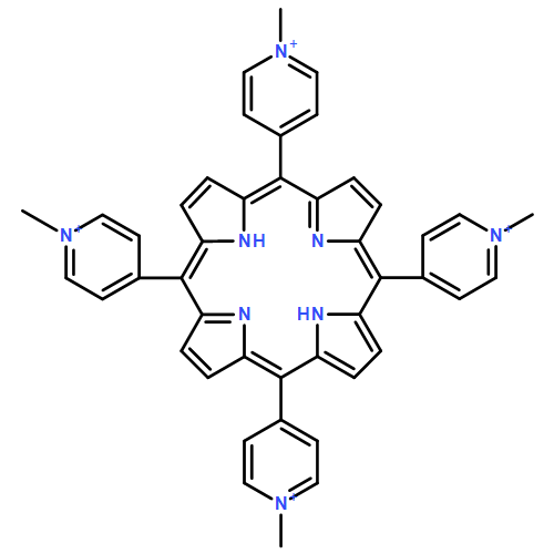
To improve the photodynamic detection and therapy of cancers (PDT), folic acid (FA) was conjugated with zinc tetraaminophthalocyanine (ZnaPc) to form ZnaPc–FA. The uptake efficiency of ZnaPc–FA to a FR-positive (folate receptor overexpressed) KB cell line (human nasopharyngeal epidermal carcinoma) was much higher than that of ZnaPc demonstrating an enhanced binding ability of ZnaPc–FA to KB cells. When KB cells were pretreated with free FA followed by incubation of ZnaPc–FA, the high uptake rate of ZnaPc–FA disappeared which demonstrated the special binding function of the FA terminal of ZnaPc–FA on KB cells. The confocal fluorescence images further showed that the affinity of ZnaPc–FA to FR-negative A549 cells (human lung epithelial carcinoma cancer cells) was very low, confirming that ZnaPc–FA can only target FR-positive cancers. The two-photon absorption cross-section of ZnaPc–FA was also higher than that of sulfonated aluminum phthalocyanine (AlPcS), an approved PS for clinical applications. With a 780 nm femto-second (fs) laser, the fluorescence image of ZnaPc–FA in KB cells under two-photon excitation (TPE) can be clearly seen, and the two-photon induced singlet oxygen in ZnaPc–FA solution was found to be proportional to the irradiation dose of the fs laser. The PDT damaging effect of ZnaPc–FA on KB cells was much effective relative to AlPcS under common one-photon excitation, and the killing efficacy of ZnaPc–FA under TPE was 10-fold higher than that of AlPcS. These results suggest that ZnaPc–FA is a promising candidate for PDT improvements and particularly for TPE PDT.
Co-reporter:Yong-Kui Xu, Sekyu Hwang, Sungjee Kim, and Ji-Yao Chen
ACS Applied Materials & Interfaces 2014 Volume 6(Issue 8) pp:5619
Publication Date(Web):March 24, 2014
DOI:10.1021/am500106c
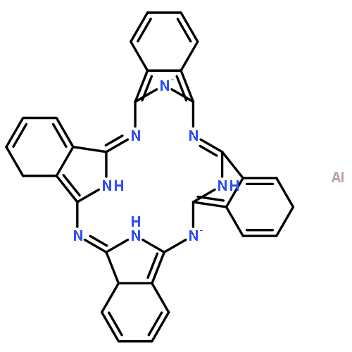
The metal-enhanced fluorescence (MEF) by metal nanoparticles is a useful technique for fluorescence detections in biological systems. The MEF effects with gold nanorods (AuNRs) and nanocubes (AuNCs) for fluorescence enhancements of sulfonated aluminum phthalocyanine (AlPcS), a commonly used and clinical approved photosensitizer for photodynamic therapy of cancers, were studied in this work. For the AuNRs which have the low aspect ratios with the corresponding longitudinal surface plasma resonance (LSPR) band in the region of 600–750 nm, the fluorescence quenching of conjugated AlPcS was found. Whereas for the AuNRs that have the LSPR bands of 800–900 nm, the MEF of AlPcS was obtained with the enhancing factor of 2–6 times, respectively. Using AuNCs, a great enhancement of AlPcS fluorescence was achieved with an enhancing factor of 150 times. Using two cancer cell lines as in vitro models, an outstanding fluorescence enhancement of AlPcS-AuNCs conjugates in cells, relative to AlPcS alone, was obtained under one-photon excitation (OPE) of 405 nm. Moreover, the bright fluorescence image of AlPcS-AuNCs in cells was also achieved under the two-photon excitation (TPE) of an 800 nm femtosecond laser. The high-quality cell imaging with either OPE or TPE demonstrated the potential of AlPcS-AuNCs in cancer cell detections.Keywords: aluminum phthalocyanine; gold nanocubes; gold nanorods; metal-enhanced fluorescence; surface plasma resonance;
Co-reporter:Jing Wang, Zehui Zhang, Shuai Zha, Yinyan Zhu, Peiyi Wu, Benjamin Ehrenberg, Ji-Yao Chen
Biomaterials 2014 35(34) pp: 9372-9381
Publication Date(Web):
DOI:10.1016/j.biomaterials.2014.07.063
Co-reporter:Yan Cen;Xiao Huang;Ren Zhang
Journal of Fluorescence 2014 Volume 24( Issue 5) pp:1481-1486
Publication Date(Web):2014 September
DOI:10.1007/s10895-014-1433-9
The photoluminescence (PL) properties of single gold nanorod (AuNR) under one-photon excitation (OPE) have been reported recently. In this work, the PL of AuNRs in aqueous solutions were studied with OPE of 514 or 633 nm to characterize the emissions of transverse and longitudinal surface Plasmon resonance (TSPR and LSPR) bands, because the AuNRs aqueous solution was frequently used in bio-medical applications. We found that under 514 nm OPE the TSPR emissions of four groups of AuNRs with different aspect ratios in aqueous solutions were all strong dominating the PL emission with the quantum yield (QY) of 10−4, which is at least three orders of magnitude higher than that of single AuNR. We further found that the aggregate was the basic form of AuNRs in aqueous solution and living cells, measured by the elastic light scattering and transmission electron microscopy measurements. The Plasmon coupling particularly the TSPR coupling between the neighbored AuNRs in aggregates enhanced the PL and increased the QY, because the conjugation of the rod side to side was a main aggregate mode. Under 633 nm OPE, only LSPR emissions of AuNRs aqueous solutions occurred with the QY level of 10−5 which is very similar to that of singe AuNR, because of the negligible LSPR coupling.
Co-reporter:Kai-Liang Chou, Nayoun Won, Jungheon Kwag, Sungjee Kim and Ji-Yao Chen
Journal of Materials Chemistry A 2013 vol. 1(Issue 36) pp:4584-4592
Publication Date(Web):04 Jul 2013
DOI:10.1039/C3TB20928H
Focusing the femto-second (fs) laser beam on the target was the usual way to carry out a two-photon excitation (TPE) in previous photodynamic therapy (PDT) studies. However, focusing the laser deep inside the tissues of the tumor is unrealistic due to tissue scattering, so that this focusing manner seems unfit for practical TPE PDT applications. In this work, we prepared a conjugate of quantum dots (QDs) and sulfonated aluminum phthalocyanine (AlPcS) for TPE PDT, because QDs have a very high two-photon absorption cross section (TPACS) and thus QDs can be excited by an unfocused 800 nm fs laser beam with a low power density and then transfer the energy to a conjugated AlPcS via fluorescence resonance energy transfer (FRET). The FRET efficiency of the QD–AlPcS conjugate in water was as high as 90%, and the FRET process of the cellular QD–AlPcS was also observed in both KB and HeLa cells under TPE of a 800 nm fs laser. The singlet oxygen (1O2) products were produced by the QD–AlPcS under the TPE of the unfocused 800 nm fs laser via FRET mediated PDT. Moreover, the QD–AlPcS can effectively destroy these cancer cells under the irradiation of the 800 nm unfocused fs laser beam with a power density of 92 mW mm−2, and particularly the killing efficiency of the TPE is comparable to that of the commonly used one-photon excitation (OPE) at visible wavelengths. These results highlight the potential of QD–AlPcS for TPE PDT with a near infrared wavelength.
Co-reporter:Xiao Huang, Xue-Jiao Tian, Wu-li Yang, Benjamin Ehrenberg and Ji-Yao Chen
Physical Chemistry Chemical Physics 2013 vol. 15(Issue 38) pp:15727-15733
Publication Date(Web):19 Mar 2013
DOI:10.1039/C3CP44227F
Gold nanorods (AuNRs) were conjugated with chlorin e6 (Ce6), a commonly used photosensitizer, to form AuNRs–Ce6 by electrostatic binding. Due to the strong surface plasmon resonance coupling, the fluorescence of conjugated Ce6 was enhanced 3-fold and the production of singlet oxygen was increased 1.4-fold. AuNRs–Ce6 were taken up by the HeLa and KB cell lines more easily than free Ce6, enhancing the intracellular delivery of Ce6. The increased cellular amount of Ce6 leads to a 3-fold more efficient photodynamic killing of these two cell lines. This demonstrates the potential of this approach to improve photodynamic detection and therapy of cancers.
Co-reporter:Xi Wu, Ji-Yao Chen, Andreas Brech, Caihong Fang, Jianfang Wang, P. Johannes Helm, Qian Peng
Biomaterials 2013 34(26) pp: 6157-6162
Publication Date(Web):
DOI:10.1016/j.biomaterials.2013.04.048
Co-reporter:X. X. Yu, J. N. Li, K. C. Kwok, M. C. Paau, Martin M. F. Choi, K. K. Shiu, J. Y. Chen, and N. H. Cheung
The Journal of Physical Chemistry C 2012 Volume 116(Issue 34) pp:18479-18486
Publication Date(Web):August 2, 2012
DOI:10.1021/jp304494t
Commercial CdSe–ZnS–polymer quantum dots (QDs) were shown to be readily quenched by strong oxidants such as potassium permanganate. When exposed to permanganate solution of millimolar concentration for hours, damage was severe and the QDs would be permanently bleached. When oxidant concentration was micromolar and reaction time was minutes, oxidation would be mild and bleached QDs could recover their fluorescence if the oxidant was removed and the QDs subsequently irradiated. Light was essential in activating the fluorescence recovery. Light could also transform mild oxidations into severe ones if administered with the oxidant simultaneously. A model of the bleaching and recovery was proposed. It was based on the oxidative attack of the Zn–ligand bond and its repair. The model helped explain the critical role that light played and was consistent with all the experimental observations. By means of photocatalyzed oxidation of single QDs, a redox probe of high spatial and temporal resolution was demonstrated.
Co-reporter:Qi-Feng Ma, Ji-Yao Chen, Xi wu, Pei-Nan Wang, Yang Yue, Ning Dai
Journal of Luminescence 2011 Volume 131(Issue 11) pp:2267-2272
Publication Date(Web):November 2011
DOI:10.1016/j.jlumin.2011.05.055
The photostability is an outstanding feature of quantum dots (QDs) used as fluorescence probes in biological staining and cell imaging. To find out the related factors in the QD photostability, the photobleaching of naked CdTe QDs and BSA coated CdSe/CdS/ZnS QDs in human hepatocellular carcinoma (QGY) cells and human nasopharynx carcinoma (KB) cells were studied under single photon excitation (SPE) and two-photon excitation (TPE). In these two cell lines the cellular QDs were irradiated by a 405 nm continuous wave laser for SPE or an 800 nm femto-second (fs) laser for TPE. The QD photobleaching with the irradiation time was found to fit a biexponential decay. The fast decay plays a dominant role in the bleaching course and thus can be used as the parameter to quantitatively evaluate the QD photostability. The TPE decreased the QD photobleaching as compared to SPE. The BSA coated core/shell QDs had improved the photostability up to 4–5 times than the naked QDs due to the shielding effect of the QD shell. Therefore, it is better to use core/shell structured QDs as the fluorescence probe combining with a TPE manner for those long-term monitoring studies.Highlights► Core/shell QDs improve the photostability up to 4–5 times as compared to the naked QDs. ► TPE is better than SPE on decreasing the photobleaching of cellular QDs. ► Combination of core/shell QDs with the TPE has the advantage for long-term cell imaging.
Co-reporter:He Meng;Lan Mi;Pei-Nan Wang
JBIC Journal of Biological Inorganic Chemistry 2011 Volume 16( Issue 1) pp:117-123
Publication Date(Web):2011 January
DOI:10.1007/s00775-010-0709-z
Bovine serum albumin (BSA)-coated CdTe/ZnS quantum dots (BSA–QDs) were selected to conjugate with folic acid (FA), forming FA–BSA–QDs. This study aims to develop these small FA–BSA–QDs (less than 10 nm) for the diagnosis of cancers in which the FA receptor (FR) is overexpressed. The enhancement of cellular uptake in FR-positive human nasopharyngeal carcinoma cells (KB cells) for FA–BSA–QDs was found by means of confocal fluorescence microscopy under single-photon and two-photon excitation. The uptake enhancement for FA–BSA–QDs was further evaluated by flow-cytometric analysis in 104 KB cells, and was about 3 times higher than for BSA–QDs on average. The uptake enhancement was suppressed when KB cells had been pretreated with excess FA, reflecting that the enhancement was mediated by the association of FR at cell membranes with FA–BSA–QDs. When human embryonic kidney cells (293T) (FR-negative cells) and KB cells, respectively, were incubated with FA–BSA–QDs (1 μM) for 40 min, the FA–BSA–QD uptake by 293T cells was much weaker than that by KB cells, demonstrating that FA–BSA–QDs could undergo preferential binding on FR-positive cancer cells. These characteristics suggest that FA–BSA–QDs are potential candidates for cancer diagnosis.
Co-reporter:Xi Wu, Tian Ming, Xin Wang, Peinan Wang, Jianfang Wang and Jiyao Chen
ACS Nano 2010 Volume 4(Issue 1) pp:113
Publication Date(Web):December 16, 2009
DOI:10.1021/nn901064m
Gold nanocubes demonstrate unique optical properties of the high photoluminescence (PL) quantum yield and a remarkably enhanced extinction band at 544 nm. The 4 × 10⁻² PL yield, which is about 200 times higher than that of gold nanorods, allows gold nanocubes to be successfully used in cell imaging of human liver cancer cells (QGY) and human embryo kidney cells (293T) with a common method of single-photon excitation. The high extinction coefficients of gold nanocubes also facilitate them carrying out the photothermal therapy of QGY and 293T cells, showing similar photokilling efficiency as compared to gold nanorods.Keywords: cell imaging; gold nanocubes; photoluminescence; photothermal therapy
Co-reporter:Lei Li, Ji-Yao Chen, Xi Wu, Pei-Nan Wang, and Qian Peng
The Journal of Physical Chemistry B 2010 Volume 114(Issue 51) pp:17194-17200
Publication Date(Web):December 7, 2010
DOI:10.1021/jp109363n
Hexadecyltrimethylammonium bromide-coated gold nanorods (AuNRs) with positive charges were effectively bound to negatively charged sulfonated aluminum phthalocyanine (AlPcS), a photosensitizer for photodynamic detection and therapy, due to the electrostatic force, with a loading content of 104 AlPcS molecules per rod. A 5-fold increase in the AlPcS fluorescence of the AlPcS−AuNRs complex was seen. The excitation fluorescence spectrum of the AlPcS−AuNRs with a typical 520 nm band fits well with the resonance band of AuNR surface plasmons, suggesting that such increased AlPcS fluorescence is produced from the strong surface plasmons of AuNRs. The intracellular distribution of AlPcS−AuNRs was studied in the QGY liver cancer cells by respectively imaging the AlPcS fluorescence and AuNRs reflectance with a confocal microscope. Furthermore, the AlPcS−AuNRs-loaded cells were photodynamically damaged after being exposed to red light in a light-dose-dependent manner. In contrast, no phototoxicity of the cells was seen after incubation with the same amount of free AlPcS, indicating that the AlPcS−AuNRs can enhance the AlPcS-mediated photodynamic effect. In addition, the loaded AlPcS can be photothermally released from AuNRs in the cells by the irradiation with an 800 nm femtosecond laser, demonstrating the potential for controlled drug release.
Co-reporter:Tao Wang;Shen Zhen;Pei-Nan Wang;Chang-Chun Wang
Journal of Fluorescence 2009 Volume 19( Issue 4) pp:615-621
Publication Date(Web):2009/07/01
DOI:10.1007/s10895-008-0452-9
To effectively image living cells with quantum dots (QDs), particularly for those cells containing high content of native fluorophores, the two-photon excitation (TPE) with a femto-second 800 nm laser was employed and compared with the single-photon excitations (SPE) of 405 nm and 488 nm in BY-2 Tobacco (BY-2-T) and human hepatocellular carcinoma (QGY) cells, respectively. The 405 nm SPE produced the bright photoluminescence (PL) signals of cellular QDs but also induced a strong autofluorescence(AF) from the native fluorophores like flavins in cells. The AF occupied about 30% and 13% of the total signals detected in QD imaging channel in the BY-2-T and QGY cells, respectively. With the excitation of 488 nm SPE, the PL signals were lower than those excited with the 405 nm SPE, although the AF signals were also reduced. The 800 nm TPE generated the best PL images of intracellular QDs with the highest signal ratio of PL to AF, because the two-photon absorption cross section of QDs is much higher than that of the native fluorophores. By means of the TPE, the reliable cellular imaging with QDs, even for the cells having the high AF background, can be achieved.
Co-reporter:Yu Zhang, Lan Mi, Pei-Nan Wang, Jiong Ma, Ji-Yao Chen
Journal of Luminescence 2008 Volume 128(Issue 12) pp:1948-1951
Publication Date(Web):December 2008
DOI:10.1016/j.jlumin.2008.06.004
pH-dependent aggregation of thiol-capped CdTe quantum dots (QDs) in solutions was observed with a confocal microscope. The average size of the QD aggregates increased from 28 nm to 1.4 μm as the pH decreased from 12 to 3. The basic condition improved the dispersion of QDs while the acidic condition caused the detachment of surface ligands, leading to the aggregation of QDs. A PL lifetime of 80 ns was detected for QDs at pH from 12 to 7, while it was shortened to 57 and 34 ns at pH 5 and 3, respectively, due to the formation of surface defects.
Co-reporter:Tian Ming;Xiaoshan Kou;Huanjun Chen;Tao Wang;Hoi-Lam Tam Dr.;Kok-Wai Cheah ;Jianfang Wang
Angewandte Chemie 2008 Volume 120( Issue 50) pp:9831-9836
Publication Date(Web):
DOI:10.1002/ange.200803642
Co-reporter:Tian Ming;Xiaoshan Kou;Huanjun Chen;Tao Wang;Hoi-Lam Tam Dr.;Kok-Wai Cheah ;Jianfang Wang
Angewandte Chemie International Edition 2008 Volume 47( Issue 50) pp:9685-9690
Publication Date(Web):
DOI:10.1002/anie.200803642
Co-reporter:Wen-Zhong Yang;Ji-Tong Yu;Lu-Wei Zhou
Photochemistry and Photobiology 2007 Volume 83(Issue 4) pp:979-984
Publication Date(Web):22 MAY 2007
DOI:10.1111/j.1751-1097.2007.00116.x
Although laser irradiation has been reported to promote skin wound healing, the mechanism is still unclear. As mast cells are found to accumulate at the site of skin wounds we hypothesized that mast cells might be involved in the biological effects of laser irradiation. In this work the mast cells, RBL-2H3, were used in vitro to investigate the effects of laser irradiation on cellular responses. After laser irradiation, the amount of intracellular calcium ([Ca2+]i) was increased, followed by histamine release, as measured by confocal fluorescence microscopy with Fluo-3/AM staining and a fluorescence spectrometer with o-phthalaldehyde staining, respectively. The histamine release was mediated by the increment of [Ca2+]i from the influx of the extracellular buffer solution through the cation channel protein, transient receptor potential vanilloid 4 (TRPV4). The TRPV4 inhibitor, Ruthenium Red (RR) can effectively block such histamine release, indicating that TRPV4 was the key factor responding to laser irradiation. These induced responses of mast cells may provide an explanation for the biological effects of laser irradiation on promoting wound healing, as histamine is known to have multi-functions on accelerating wound healing.
Co-reporter:F. L. Xue;J. Y. Chen;J. Guo;C. C. Wang;W. L. Yang
Journal of Fluorescence 2007 Volume 17( Issue 2) pp:149-154
Publication Date(Web):2007 March
DOI:10.1007/s10895-006-0152-2
Quantum dots (QDs), as novel fluorescence probes, have shown a great potential for bio-molecular labeling and cellular imaging. To stain cellular targets, the sufficient intracellular delivery of QDs is required. In this work the tat, a typical membrane-permeable carrier peptide, was conjugated with thiol-capped CdTe QDs to form CdTe Tat-QDs, and the intracellular deliveries of CdTe QDs or CdTe Tat-QDs were compared in human hepatocellular carcinoma (QGY) cells and human breast cancer (MCF7) cells in vitro by means of confocal laser scanning microscopy. Added into the cell dishes, both QDs and Tat-QDs adhered to the outer leaflet of the plasma membrane of cells within a few minutes, but the binding amount of Tat-QDs was obviously higher than that of QDs. Then both QDs and Tat-QDs can penetrate into cells, and their cellular contents increased with incubation time but both saturated after 3 hours incubation. However the cellular levels of Tat-QDs were higher than those of QDs, with the ratio of 2.1 (±0.3) times in QGY cells and 1.5 (±0.2) times in MCF7 cells, demonstrating the enhancing effect of Tat conjugation on the intracellular delivery of QDs.
Co-reporter:X.Q. Mi, J.Y. Chen, L.W. Zhou
Journal of Photochemistry and Photobiology B: Biology 2006 Volume 83(Issue 2) pp:146-150
Publication Date(Web):1 May 2006
DOI:10.1016/j.jphotobiol.2005.12.018
In our previous study we found that low power laser irradiation improved the erythrocyte deformability, but the mechanism is unclear. The membrane-attached hemoglobin (Hbm) may be one of the determining factors for the erythrocyte deformability. We report here for the first time, that laser irradiation can reduce the Hbm contents in pig’s erythrocytes, providing the explanation for the improvement of erythrocyte deformability. The decrease of the Hbm was proportional to the irradiation dose, but the relative change of Hbm was saturated around 35%. The 532 nm laser was more efficient at lowering Hbm than the 632.8 nm laser, consistent with the absorption spectrum of Hbm.
Co-reporter:Ma Jiong;Cen Yan;Chen Ji-Yao
Chinese Journal of Chemistry 2005 Volume 23(Issue 5) pp:
Publication Date(Web):14 JUN 2005
DOI:10.1002/cjoc.200590637
The nitrosyl-hemoglobin (HbNO) is the carrier of nitric oxide (NO) which is the important messenger molecule displaying multiple physiologic and pathophysiologic roles. However it is still not clear for the fate of HbNO molecules during the venous-arterial transit. In this letter, the HbNO transition in vitro was studied by using the electron paramagnetic resonance (EPR) spectra. It was found that HbNO molecules were stable when oxygen did not exist in the system but not stable in aerobic conditions. The absorption spectra further revealed that the methemoglobin (metHb) was the product of HbNO in aerobic environment, showing that the HbNO changed to metHb when there were enough oxygen molecules in the system.
Co-reporter:X.Q Mi, J.Y Chen, Y Cen, Z.J Liang, L.W Zhou
Journal of Photochemistry and Photobiology B: Biology 2004 Volume 74(Issue 1) pp:7-12
Publication Date(Web):19 March 2004
DOI:10.1016/j.jphotobiol.2004.01.003
The effects of laser irradiation with 632.8 and 532 nm on rheological properties of blood were comparatively studied in vitro. Under the irradiation condition of 30 mW, laser irradiation of blood samples using a spot diameter of 5 mm with each laser, showed promising results in the modulation of hemorheological properties. When blood samples from patients with abnormally high values of erythrocyte sedimentation rate (ESR) were irradiated, the values of ESR were lowered statistically by either of the 632.8 or 532 nm lasers. The laser irradiation reduced blood viscosities at different shear rates (10–110 S−1) for the hyper-viscosity blood samples. Laser irradiation increased the electrophoretic mobility (EPM) of erythrocytes when the values of the sample's EPM were abnormally slow. The erythrocyte deformability was enhanced by laser irradiation when the deformability of the sample from the patients was originally poor. For verifying the improvement of laser irradiation on erythrocyte deformability, the typical erythrocyte samples with poor deformability were produced by the pre-treatment of the erythrocytes with Ca2+. The deformability of these erythrocyte samples was also improved after laser irradiation. These results suggest that membrane-bound hemoglobin (Hbm) might be the initial site of the interaction, since Hbm is the main cause of poor deformability when erythrocytes were treated with Ca2+. In all experiments including ESR, blood viscosity, EPM and erythrocyte deformability, the 532 nm laser demonstrated more efficient effects on modulating rheological properties than 632.8 nm laser. This wavelength effect is consistent with the absorption spectrum of hemoglobin, reflecting that hemoglobin may be one of the action targets under laser irradiation.
Co-reporter:J.Y. Chen, Q. Peng, H.-J. Jodl
Spectrochimica Acta Part A: Molecular and Biomolecular Spectroscopy 2003 Volume 59(Issue 11) pp:2571-2576
Publication Date(Web):September 2003
DOI:10.1016/S1386-1425(03)00011-8
5-Aminolevulinic acid (ALA) and its hexyl ester (ALA-H) are bioorganic molecules, now used as drugs in the study and clinical application of photodynamic therapy (PDT). Their infrared spectra were reported in first time here. The spectral characteristic was found well correlated to their structure feature. The strong peaks of CO, CH2 and OH band were shown in ALA spectrum. While in case of ALA-H, besides the vibration modes of CO and CH2 the additional CH3 infrared peaks appeared, which correspond with their structural difference. Thus the infrared spectrum could be used to detect and distinguish ALA and ALA-H, which have potential for the mechanism study of ALA and ALA-H based PDT in biological system. Using the infrared spectrum as the probe, the thermal effect on structure stability was detected. Below the temperature of 80 °C, the ALA and ALA-H are thermally stable in structure. When temperature reached 120 °C, the serious structure breaking (thermal decomposition) happened for both ALA and ALA-H.
Co-reporter:Jing Wang, Dai-Xin Ye, Guo-Hai Liang, Jian Chang, Ji-Lie Kong and Ji-Yao Chen
Journal of Materials Chemistry A 2014 - vol. 2(Issue 27) pp:NaN4345-4345
Publication Date(Web):2014/05/02
DOI:10.1039/C4TB00366G
We report a novel method for synthesizing water-dispersible silicon nanoparticles (Si NPs) with a simple one-step procedure using mild reagents (3-aminopropyl) trimethoxysilane (APTES) and ascorbate sodium (AS). This is the first report of “green” synthesis of Si NPs on a large scale and at low cost. The method involves a quick reaction in a commonly used round bottom flask at room temperature and pressure without additional treatment and any special equipment. The as-prepared Si NPs have an average diameter of 2 nm and an emission band at 530 nm with a full width at half maximum height (FWHM) of 70 nm and a quantum yield (QY) of 0.21. Moreover, the fluorescence lifetime of these Si NPs is much longer than that of native fluorophores in living cells. Therefore, these Si NPs allow effective imaging of living cells with a fluorescence lifetime imaging microscope (FLIM). Using the time gating model in FLIM, an excellent image was obtained in which the auto-fluorescence interference of cellular fluorophores was suppressed demonstrating that the Si NPs are promising probes for cell imaging particularly using the FLIM technique.
Co-reporter:Xiao Huang, Xue-Jiao Tian, Wu-li Yang, Benjamin Ehrenberg and Ji-Yao Chen
Physical Chemistry Chemical Physics 2013 - vol. 15(Issue 38) pp:NaN15733-15733
Publication Date(Web):2013/03/19
DOI:10.1039/C3CP44227F
Gold nanorods (AuNRs) were conjugated with chlorin e6 (Ce6), a commonly used photosensitizer, to form AuNRs–Ce6 by electrostatic binding. Due to the strong surface plasmon resonance coupling, the fluorescence of conjugated Ce6 was enhanced 3-fold and the production of singlet oxygen was increased 1.4-fold. AuNRs–Ce6 were taken up by the HeLa and KB cell lines more easily than free Ce6, enhancing the intracellular delivery of Ce6. The increased cellular amount of Ce6 leads to a 3-fold more efficient photodynamic killing of these two cell lines. This demonstrates the potential of this approach to improve photodynamic detection and therapy of cancers.
Co-reporter:By Song Wang, Jing Wang and Ji-Yao Chen
Journal of Materials Chemistry A 2014 - vol. 2(Issue 11) pp:NaN1602-1602
Publication Date(Web):2014/01/09
DOI:10.1039/C3TB21620A
To improve the photodynamic detection and therapy of cancers (PDT), folic acid (FA) was conjugated with zinc tetraaminophthalocyanine (ZnaPc) to form ZnaPc–FA. The uptake efficiency of ZnaPc–FA to a FR-positive (folate receptor overexpressed) KB cell line (human nasopharyngeal epidermal carcinoma) was much higher than that of ZnaPc demonstrating an enhanced binding ability of ZnaPc–FA to KB cells. When KB cells were pretreated with free FA followed by incubation of ZnaPc–FA, the high uptake rate of ZnaPc–FA disappeared which demonstrated the special binding function of the FA terminal of ZnaPc–FA on KB cells. The confocal fluorescence images further showed that the affinity of ZnaPc–FA to FR-negative A549 cells (human lung epithelial carcinoma cancer cells) was very low, confirming that ZnaPc–FA can only target FR-positive cancers. The two-photon absorption cross-section of ZnaPc–FA was also higher than that of sulfonated aluminum phthalocyanine (AlPcS), an approved PS for clinical applications. With a 780 nm femto-second (fs) laser, the fluorescence image of ZnaPc–FA in KB cells under two-photon excitation (TPE) can be clearly seen, and the two-photon induced singlet oxygen in ZnaPc–FA solution was found to be proportional to the irradiation dose of the fs laser. The PDT damaging effect of ZnaPc–FA on KB cells was much effective relative to AlPcS under common one-photon excitation, and the killing efficacy of ZnaPc–FA under TPE was 10-fold higher than that of AlPcS. These results suggest that ZnaPc–FA is a promising candidate for PDT improvements and particularly for TPE PDT.
Co-reporter:Kai-Liang Chou, Nayoun Won, Jungheon Kwag, Sungjee Kim and Ji-Yao Chen
Journal of Materials Chemistry A 2013 - vol. 1(Issue 36) pp:NaN4592-4592
Publication Date(Web):2013/07/04
DOI:10.1039/C3TB20928H
Focusing the femto-second (fs) laser beam on the target was the usual way to carry out a two-photon excitation (TPE) in previous photodynamic therapy (PDT) studies. However, focusing the laser deep inside the tissues of the tumor is unrealistic due to tissue scattering, so that this focusing manner seems unfit for practical TPE PDT applications. In this work, we prepared a conjugate of quantum dots (QDs) and sulfonated aluminum phthalocyanine (AlPcS) for TPE PDT, because QDs have a very high two-photon absorption cross section (TPACS) and thus QDs can be excited by an unfocused 800 nm fs laser beam with a low power density and then transfer the energy to a conjugated AlPcS via fluorescence resonance energy transfer (FRET). The FRET efficiency of the QD–AlPcS conjugate in water was as high as 90%, and the FRET process of the cellular QD–AlPcS was also observed in both KB and HeLa cells under TPE of a 800 nm fs laser. The singlet oxygen (1O2) products were produced by the QD–AlPcS under the TPE of the unfocused 800 nm fs laser via FRET mediated PDT. Moreover, the QD–AlPcS can effectively destroy these cancer cells under the irradiation of the 800 nm unfocused fs laser beam with a power density of 92 mW mm−2, and particularly the killing efficiency of the TPE is comparable to that of the commonly used one-photon excitation (OPE) at visible wavelengths. These results highlight the potential of QD–AlPcS for TPE PDT with a near infrared wavelength.