Co-reporter:Xiaojun Qu;Yuqian Liu;Qingsheng Guo;Xuebin Huang
ACS Applied Materials & Interfaces February 8, 2017 Volume 9(Issue 5) pp:4725-4732
Publication Date(Web):January 13, 2017
DOI:10.1021/acsami.6b14972
In this work, we report a new type of quantum dot (QD)-based fluorescence resonance energy transfer (FRET) assembly and its utility for sensing Zn2+ in different media. The assembly on the QD scaffold is via first coating of poly(dA) homopolymer/double-stranded DNA, followed by loading of meso-tetra(4-sulfonatophenyl)porphine dihydrochloride (TSPP), both of which are electrostatic, offering the advantages of cost-efficiency and simplicity. More importantly, the biopolymer coating minimizes the interfacial thickness to be ≤2 nm for QD-TSPP FRET, which results in improvements of up to 60-fold for single FRET efficiency and nearly 4-fold for total FRET efficiency of the QD-biopolymer-TSPP assemblies in comparison with silica-coating-based QD-TSPP assemblies. On the basis of Zn2+-chelation-induced spectral modulation, dual-emission QD-poly(dA)-TSPP assemblies are developed as a ratiometric Zn2+ sensor with increased sensitivity and specificity. The sensor either in solution or on a paper substrate displays continuous color changes from yellow to bright green toward Zn2+, exhibiting excellent visualization capability. By utilizing the competitive displacement of Zn2+, the sensor is also demonstrated to have good reversibility. Furthermore, the sensor is successfully used to visualize exogenous Zn2+ in living cells. Together the QD-biopolymer-TSPP assembly provides an inexpensive, sensitive, and reliable sensing platform not only for on-site analytical applications but also for high-resolution cellular imaging.Keywords: biopolymer; electrostatic assembly; porphyrin; quantum dot; resonance energy transfer; sensor; zinc ion;
Co-reporter:Xianyun Hu;Yuqian Liu;Xiaojun Qu
Chemical Communications 2017 vol. 53(Issue 81) pp:11181-11184
Publication Date(Web):2017/10/10
DOI:10.1039/C7CC05279K
By a combination of quantum dot-labelled aptamers and graphene oxide, a hybrid molecular system was developed for the integration of multiple logic gates to implement half adder and half subtractor functions. On the merits of quantum dots, repetitious arithmetic operations and a reliable fluorescent switch were demonstrated.
Co-reporter:Qingsheng Guo;Feika Bian;Yuqian Liu;Xiaojun Qu;Xianyun Hu
Chemical Communications 2017 vol. 53(Issue 36) pp:4954-4957
Publication Date(Web):2017/05/02
DOI:10.1039/C7CC00462A
A robust Qbead platform that integrated with target-binding, hybridization chain reaction and staining was developed for the direct multiplexed detection of endogenous miRNAs by amplified Qbead-colour change.
Co-reporter:Haojun Jin, Yuqian Liu, Tianshu Xu, Xiaojun Qu, Feika Bian, and Qingjiang Sun
Analytical Chemistry 2016 Volume 88(Issue 21) pp:10411
Publication Date(Web):October 1, 2016
DOI:10.1021/acs.analchem.6b01967
By complexing a nonionic G-quadruplex ligand with hybrid dual-emission quantum dots (QDs), a ratiometric fluorescent nanoprobe is developed for G-quadruplex detection in a sensitive and specific manner. The QDs nanohybrid comprised of a green-emission QD (gQD) and multiple red-emission QDs (rQDs) inside and outside of a silica shell, respectively, is utilized as the signal displaying unit. Only the presence of G-quadruplex can displace the ligand from QDs, breaking up the QDs–ligand complexation, and inducing the restoration of the rQDs fluorescence. Since the fluorescence of embedded gQD stays constant, variations of the dual-emission intensity ratios display continuous color changes from green to bright orange, which can be clearly observed by the naked eye. Furthermore, by utilizing competitive binding of a cationic ligand versus the nonionic ligand toward G-quadruplex, the nanoprobe is demonstrated to be applicable for assessing the affinity of a G-quadruplex-targeted anticancer drug candidate, exhibiting ratiometric fluorescence signals (reverse of that for G-quadruplex detection). By making use of the specificity of the ligand binding with G-quadruplex against a double helix, this nanoprobe is also demonstrated to be capable of sensitive detection of one-base mutation, exhibiting sequence-specific ratiometric fluorescence signals. By functionalizing with a nuclear localization peptide, the nanoprobe can be used for visualization of G-quadruplex in the nucleus of human cells.
Co-reporter:Yuqian Liu, Mingfu Ye, Qinyu Ge, Xiaojun Qu, Qingsheng Guo, Xianyun Hu, and Qingjiang Sun
Analytical Chemistry 2016 Volume 88(Issue 3) pp:1768
Publication Date(Web):January 11, 2016
DOI:10.1021/acs.analchem.5b04043
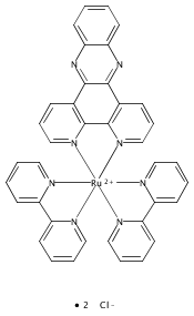
We have developed a proof-of-concept quantum dot–ligand (QD–L) system for visual selective detection of nucleic acids, in combination with a ratiometric fluorescence technique. This system comprises a dual-emission QDs nanohybrid formed by embedding a red-emission QD (rQD) in a silica nanoparticle and electrostatically assembling green-emission QDs (gQDs) onto the silica surface, as the signal displaying unit, and a hydrophobic compound, dipyrido[3,2-a:2′,3′-c]phenazine (dppz), attached onto the gQDs surface via phase transfer, as the ligand as well as fluorescence quencher of gQDs. This system is successfully used for quantification of double-stranded DNA (dsDNA). Because of its avid binding with dppz, dsDNA can break up the QD–L system, displacing the dppz ligand from the gQDs surface and restoring the gQDs emission. Since the red emission of embedded rQDs stays constant, variations of the dual-emission intensity ratios display continuous color changes from orange to bright green, which can be clearly observed by the naked eye. More importantly, this system is advantageous in terms of specificity over a QD ionic conjugate, because the electrical neutrality of dppz excludes its nonspecific electrostatic association with dsDNA. The QD–L system also is capable of detecting single-nucleotide polymorphism, exhibiting sequence-specific ratiometric fluorescence as a QD-bioconjugate does, but possessing the obvious advantage in terms of low cost, with the avoidance of modification, labeling, and purification processes. Therefore, the QD–L system provides an extremely simple but general strategy for detecting nucleic acids in a facile, sensitive, and specific manner.
Co-reporter:Qingsheng Guo, Zhixiong Bai, Yuqian Liu, Qingjiang Sun
Biosensors and Bioelectronics 2016 Volume 77() pp:107-110
Publication Date(Web):15 March 2016
DOI:10.1016/j.bios.2015.09.031
•A microarray assay is demonstrated with streptavidin coated quantum dot labeling immobilized molecular beacon.•With the regeneration of molecular beacon, the microarray can be reused, lowering the assay cost.•The microarray is successfully used to detect single nucleotide polymorphisms.•The microarray can perform as a 4-to-2 encoder.In this work, we report the application of streptavidin-coated quantum dot (strAV-QD) in molecular beacon (MB) microarray assays by using the strAV-QD to label the immobilized MB, avoiding target labeling and meanwhile obviating the use of amplification. The MBs are stem-loop structured oligodeoxynucleotides, modified with a thiol and a biotin at two terminals of the stem. With the strAV-QD labeling an “opened” MB rather than a “closed” MB via streptavidin-biotin reaction, a sensitive and specific detection of label-free target DNA sequence is demonstrated by the MB microarray, with a signal-to-background ratio of 8. The immobilized MBs can be perfectly regenerated, allowing the reuse of the microarray. The MB microarray also is able to detect single nucleotide polymorphisms, exhibiting genotype-dependent fluorescence signals. It is demonstrated that the MB microarray can perform as a 4-to-2 encoder, compressing the genotype information into two outputs.
Co-reporter:Liang Wu, Qing-Sheng Guo, Yu-Qian Liu, and Qing-Jiang Sun
Analytical Chemistry 2015 Volume 87(Issue 10) pp:5318
Publication Date(Web):May 1, 2015
DOI:10.1021/acs.analchem.5b00514
In this work, we report the design and application of a new ratiometric fluorescent probe, which contains different-colored quantum dots (QDs) as dual fluorophores, ultrathin silica shell as spacer, and meso-tetra(4-sulfonatophenyl)porphine dihydrochloride (TSPP) as receptor, for Zn2+ detection in aqueous solution and living cells. In the architecture of our designed probe, the silica shell plays the key roles in controlling the locations of QDs, TSPP, and Zn2+, preventing the direct contact between QDs and Zn2+ but affording fluorescence resonance energy transfer (FRET) from dual-color QDs to TSPP. In the presence of Zn2+, the analyte-receptor reaction changes the absorption in the range of the Q-band of TSPP and accordingly the efficiencies of two independent FRET processes from the dual-colored QDs to the acceptor, respectively, leading to fluorescence enhancement of green-emission QDs whereas fluorescence quenching of yellow-emission QDs. Benefiting from the well-resolved dual emissions from different-colored QDs and the large range of emission ratios, the probe solution displays continuous color changes from yellow to green, which can be clearly observed by the naked eye. Under physiological conditions, the probe exhibits a stable response for Zn2+ from 0.3 to 6 μM, with a detection limit of 60 nM in aqueous solutions. With respect to single-emission probes, this ratiometric probe has demonstrated to feature excellent selectivity for Zn2+ over other physiologically important cations such as Fe3+ and Cu2+. It has been preliminarily used for ratiometric imaging of Zn2+ in living cells with satisfying resolution.
Co-reporter:Kao Zhu, Xianyun Hu, Qinyu Ge, Qingjiang Sun
Analytica Chimica Acta 2014 Volume 812() pp:199-205
Publication Date(Web):17 February 2014
DOI:10.1016/j.aca.2014.01.007
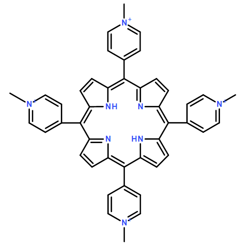
•The QD/TMPyP complex made by electrostatic association is used for fluorescent detection of DNAs.•The detection limit of 0.16 nM and a linear range of 0.25–6.0 nM are achieved.•This probe is sensitive to the binding modes of TMPyP with DNAs.•The thymine bases preferential of TMPyP–DNA interaction is proved by this probe.We have developed a new fluorescent probe of thioglycolic acid (TGA)-capped CdTe quantum dots (QDs) complexed with a model drug, meso-tetrakis(N-methylpyridinium-4-yl)porphyrin (TMPyP) for detecting deoxyribonucleic acids (DNAs). This probe operates with an “Off–On” mode: TMPyP quenches the photoluminescence (PL) of QDs via a photo induced electron-transfer (PIET) process; the presence of DNA can break the QD/TMPyP complexation, interrupting the PIET process, and switch on the PL of QDs. Sensitive detection of DNA with the detection limit of 0.16 nM and a linear detection range of 0.25–6.0 nM are achieved. Importantly, this probe can be used to distinguish the binding modes of DNA–TMPyP interactions, exhibiting the DNA sequence-dependent PL recovery behaviors. The obtained binding constant for poly(dA)·poly(dT) is ∼3.30 × 10⁷ L mol−1, which is approximately one order of magnitude larger than those for native DNAs and poly(dG)·poly(dC). Furthermore, the thymine bases preferential of the TMPyP–DNA interaction is proved by this probe.
Co-reporter:Xianyun Hu, Kao Zhu, Qingsheng Guo, Yuqian Liu, Mingfu Ye, Qingjiang Sun
Analytica Chimica Acta 2014 Volume 812() pp:191-198
Publication Date(Web):17 February 2014
DOI:10.1016/j.aca.2014.01.006
•A simple CdTe QD–Phen sensor is constructed for detecting Cd2+.•The sensor is operated with a ligand-displacement induced PL switch strategy.•The detection limit of 0.01 nM for Cd2+ is achieved.•This sensor features to discriminate Cd2+ versus Zn2+, and succeeds in real water samples.This paper reports the construction of a simple CdTe quantum dots (QDs)-based sensor with 1,10-phenanthroline (Phen) as ligand, and the demonstration of a novel ligand displacement-induced fluorescence switch strategy for sensitive and selective detection of Cd2+ in aqueous phase. The complexation of Phen at the surface quenches the green photoluminescence (PL) of QDs dominated by a photoinduced hole transfer (PHT) mechanism. In the presence of Cd2+, the Phen ligands are readily detached from the surface of CdTe QDs, forming [Cd(Phen)2(H2O)2]2+ in solution, and as a consequence the PL of CdTe QDs switches on. The detection limit for Cd2+ is defined as ∼0.01 nM, which is far below the maximum Cd2+ residue limit of drinking water allowed by the U.S. Environmental Protection Agency (EPA). Two consecutive linear ranges allow a wide determination of Cd2+ from 0.02 nM to 0.6 μM. Importantly, this CdTe QDs-based sensor features to distinctly discriminate between Cd2+ and Zn2+, and succeeds in real water samples. This extremely simple strategy reported here represents an attempt for the development of fluorescent sensors for ultrasensitive chemo/biodetection.
Co-reporter:Lei Gao;Jing Zhang;Chang He;Yi Zhang
Science China Chemistry 2014 Volume 57( Issue 7) pp:966-972
Publication Date(Web):2014 July
DOI:10.1007/s11426-014-5114-y
Photovoltaic performance of the organic solar cells (OSCs) based on 2-((5′-(4-((4-((E)-2-(5′-(2,2-dicyanovinyl)-3′,4-dihexyl-2,2′-bithiophen-5-yl)vinyl) phenyl)(phenyl)amino)styryl)-4,4′-dihexyl-2,2′-bithiophen-5-yl)methylene)malononitrile (L(TPA-bTV-DCN)) as donor and PC70BM as acceptor was optimized using 0.25 vol% high boiling point solvent additive of 1-chloronaphthalene (CN), 1,6-hexanedithiol (HDT), or 1,8-diodooctane (DIO). The optimized OSC based on L(TPA-bTV-DCN)-PC70BM (1:2, w/w) with 0.25 vol% CN exhibits an enhanced power conversion efficiency (PCE) of 2.61%, with Voc of 0.87 V, Jsc of 6.95 mA/cm2, and FF of 43.2%, under the illumination of 100 mW/cm2 AM 1.5 G simulated solar light, whereas the PCE of the OSC based on the same active layer without additive is only 1.79%. The effect of the additive on absorption spectra and the atomic force microscopy images of L(TPA-bTV-DCN)-PC70BM blend films were further investigated. The improved efficiency of the device could be ascribed to the enhanced absorption and optimized domain size in the L(TPA-bTV-DCN)-PC70BM blend film.
Co-reporter:Lu Zhang, Kao Zhu, Tao Ding, Xianyun Hu, Qingjiang Sun and Chunxiang Xu
Analyst 2013 vol. 138(Issue 3) pp:887-893
Publication Date(Web):22 Nov 2012
DOI:10.1039/C2AN36303H
We have developed a new fluorescent probe based on direct conjugation between 1,10-phenanthroline (Phen) and water-soluble thioglycolic acid (TGA) capped CdTe quantum dots (QDs) for the detection of double-stranded DNA (dsDNA). Phen could directly adsorb onto the QDs surface by metal-affinity driven coordination, quenching the photoluminescence (PL) of QDs via the photoinduced hole transfer process; addition of dsDNA would bring the restoration of QDs PL, as Phen could intercalate into dsDNA followed by its dissociation from the QDs surface. The dependence of QDs PL on the dsDNA amount as well as temperature was utilized to investigate the Phen–dsDNA interaction. The obtained binding constant of the QD–Phen dyad was 2–3 orders of magnitude higher than that of Phen-based metal complexes. Both the binding constant and the binding site of dsDNA with Phen increased with the elevated temperature, owing to an endothermic process. At 37 °C, sensitive detection of dsDNA with a detection limit of ∼3 nmol L−1 was achieved. Therefore, the QD-molecule direct conjugation based fluorescent probe could provide an effective alternative to those based on QD-bioconjugation and QD-ionic conjugation.
Co-reporter:Qingsheng Guo, Feika Bian, Yuqian Liu, Xiaojun Qu, Xianyun Hu and Qingjiang Sun
Chemical Communications 2017 - vol. 53(Issue 36) pp:NaN4957-4957
Publication Date(Web):2017/04/10
DOI:10.1039/C7CC00462A
A robust Qbead platform that integrated with target-binding, hybridization chain reaction and staining was developed for the direct multiplexed detection of endogenous miRNAs by amplified Qbead-colour change.

![Ethanol,2,2'-[[4-[[2-methoxy-5-methyl-4-[(4-methyl-2-nitrophenyl)azo]phenyl]azo]phenyl]imino]bis-](http://img.cochemist.com/ccimg/374600/374591-92-1.png)
![Ethanol,2,2'-[[4-[[2-methoxy-5-methyl-4-[(4-methyl-2-nitrophenyl)azo]phenyl]azo]phenyl]imino]bis-](http://img.cochemist.com/ccimg/374600/374591-92-1_b.png)


![Dipyrido[3,2-a:2',3'-c]phenazine](/data/chemimg/1778900/19535-47-8.png)
![Dipyrido[3,2-a:2',3'-c]phenazine](/data/chemimg/1778900/19535-47-8_b.png)