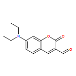
Co-reporter:Qian Sun;Weisi Wang;Zhaoyang Chen;Yuhua Yao;Weibing Zhang;Liping Duan;Junhong Qian
Chemical Communications 2017 vol. 53(Issue 48) pp:6432-6435
Publication Date(Web):2017/06/13
DOI:10.1039/C7CC03587J
A fluorescence “off–on” probe CBF, constructed by incorporating a dioxaborine unit into a microenvironment-sensitive fluorophore, was developed for serum albumin (SA). Upon binding to SA, the dioxaborine group in CBF was hydrolyzed into β-diketonate, which triggered dramatic fluorescence enhancement (over 1000-fold) along with a remarkable blue-shift (∼100 nm). The bioimaging results suggested that more SA were taken in by cancer cells.
Co-reporter:Qian Sun, Weibing Zhang, Junhong Qian
Talanta 2017 Volume 162() pp:107-113
Publication Date(Web):1 January 2017
DOI:10.1016/j.talanta.2016.10.002
•A ratiometric fluorescence probe SPH with long emission wavelength for sulfite was designed.•SPH exhibited high selectivity and sensitivity toward sulfite over other relative species.•SPH could monitor sulfite levels in realistic samples with good recovery.A new fluorescent probe 7-(diethylamino)-3-((1e,3e)-5-oxo-5-phenylpenta-1,3-dien-1-yl)-2H-chromen-2-one (SPH), based on Michael addition mechanism, was designed and synthesized for selective detection of sulfite. The probe was constructed by incorporating an α,β-unsaturated ketone conjugated with a C˭C bond into the coumarin fluorophore as a specifical reaction site for sulfite utilizing its nucleophilic property. The extra conjugated C˭C bond induced obvious red-shifts in both absorption and emission maxima, and remarkably promoted the nucleophilic addition rate. Once treated with sulfite, the solution's color changed from orange to yellow companied with a strong blue-green fluorescence. The ratio of fluorescence intensity at 488 nm and 630 nm (I488/I630) was linear with sulfite concentration over the range of 0–80 μM with a detection limit of 0.23 μM. High selectivity and good competition of the probe toward sulfite over other anions and thiols enable us to monitor sulfite levels in realistic samples as well as in living HeLa cells.A new ratiometric fluorescence probe was designed and synthesized. High selectivity and good competition of the probe ensure it a potential candidate for monitoring sulfite levels in realistic samples as well as in living Hela cells.
Co-reporter:Qian Sun, Deheng Sun, Lun Song, Zhuo Chen, Zhaoyang Chen, Weibing Zhang, and Junhong Qian
Analytical Chemistry 2016 Volume 88(Issue 6) pp:3400
Publication Date(Web):February 23, 2016
DOI:10.1021/acs.analchem.6b00178
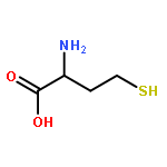
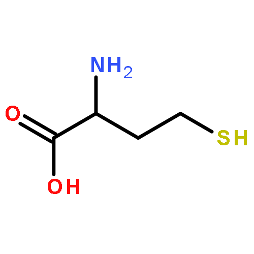
Sulfhydryl-containing proteins play critical roles in various physiological and biological processes, and the activities of those proteins have been reported to be susceptible to thiol oxidation. Therefore, the development of protein thiol target fluorescent probe is highly desirable. In the present work, a biotinylated coumarin fluorescence “off–on” probe SQ for selectively detecting protein thiols in biotin receptor-positive cancer cells was designed with a 2,4-dinitrobenzenesulfony as the thiol receptor. The probe exhibited dramatic fluorescence responses toward sulfhydryl-containing proteins (ovalbumin (OVA), bovine serum albumin (BSA)): up to 170-fold fluorescence enhancement with 70 nm blue-shift was observed with the addition of OVA. However, low molecular weight thiols (Cys, glutathione (GSH), Hcy) caused negligible fluorescence changes of SQ. In addition, biotin receptor-positive Hela cells displayed strong red and green fluorescence after incubation of SQ for 1 h; neither red nor green fluorescence signal could be visualized in biotin-negative normal lung Wi38 cells. These results imply that the probe has potential application in fluorescent imaging protein thiols on the surface of Hela cells.
Co-reporter:Lun Song, Li-Min Ma, Qian Sun, Wei-Bing Zhang, Min-Bo Lan, Jun-Hong Qian
Chinese Chemical Letters 2016 Volume 27(Issue 3) pp:330-334
Publication Date(Web):March 2016
DOI:10.1016/j.cclet.2015.12.012
4-Fluoro-7-nitrobenzo-2-oxa-1,3-diazole (NBD-F) was employed as a colorimetric probe for differential detection of biothiols in different media. The spectral response and the selectivity of NBD-F toward thiols were significantly improved by surfactant micelles. Mercapto group exhibited high reactivity in all the solvents (including Tris–HCl buffer solution, CTAB and SDS micelles). The 4-thioether derivatives of NBD-F reacting with Cys and Hcy but not GSH could transfer to the corresponding 4-amino-substituents via intramolecular nucleophilic aromatic substitution, thus, GSH could be discriminated from Cys/Hcy. In CTAB micelles, the reaction product of NBD-F with Cys is non-fluorescent and it absorbs in long-wavelength region. According to the spectral responses of NBD-F toward different low-molecular-weight thiols, we could identify Cys, Hcy and GSH from each other.4-Fluoro-7-nitrobenzo-2-oxa-1,3-diazole (NBD-F) was employed as a probe to distinguish biothiols from each other. The spectral response and the selectivity of NBD-F toward thiols were dramatically improved by surfactant micelles.
Co-reporter:Qian Sun, Haiyu Tian, Haoran Qu, Deheng Sun, Zhuo Chen, Liping Duan, Weibing Zhang and Junhong Qian
Analyst 2015 vol. 140(Issue 13) pp:4648-4653
Publication Date(Web):01 May 2015
DOI:10.1039/C5AN00585J
Two biotinylated coumarin-based fluorescent probes SPS3 and RC3 were designed for differentiating between structurally similar proteins streptavidin (SA) and avidin (AV). A substituted phenyl group is introduced onto SPS3, which may quench the fluorescence through twist intramolecular charge transfer (TICT). The fluorescence of SPS3 is turned on, by restraining the TICT process, when the fluorophore is buried at the surface of SA. RC3 is constructed by incorporating a biotin molecule to a coumarin fluorophore through a 4-atom spacer. The fluorescence intensity of RC3 is enhanced significantly when its fluorophore enters into the less polar binding pocket of AV. SPS3 and RC3 could be applied in distinguishing between SA and AV as well as in fluorescence imaging of biotin receptor over-expressed Hela cells.
Co-reporter:Shanshan Liu, Lun Song, Qian Sun, Zhaoyang Chen, Yu Ge, Weibing Zhang and Junhong Qian
RSC Advances 2015 vol. 5(Issue 111) pp:91863-91868
Publication Date(Web):22 Oct 2015
DOI:10.1039/C5RA17962A
Two colorimetric/ratiometric fluorescence probes (BSP1 and BSP2) for sulfite utilizing the Michael-addition mechanism were designed and synthesized by incorporating a substituted pyridinium/methyl pyridinium to a BODIPY fluorophore linked with an α,β-unsaturated ketone. BSP1 exhibited colorimetric and ratiometric fluorescence responses toward sulfite in PBS-CTAB system. The addition of sulfite to the CC group in BSP1 induced about 60 nm hypsochromic shift in both absorption (from 554 nm to 494 nm) and emission (from 618 nm to 554 nm) spectra accompanied with fluorescent color change from red to green under hand-lamp excited at 365 nm. Good linear relationship was found between the emission ratio (I554/I618) and the concentration of sulfite in the range of 0–300 μM with a detection limit of 6.4 μM. In the case of BSP2, the probe showed excellent water solubility and faster spectral response to sulfite with about 100 nm blue shift (from 567 nm to 470 nm) in the absorption spectrum in PBS. The solution's color changed from purple to yellow. Both BSP1 and BSP2 displayed high selectivity and competition towards sulfite over other relative species such as thiols and sulfide. More importantly, we have demonstrated that BSP1 could be applied in the imaging of sulfite in living cells.
Co-reporter:Shanshan Liu, Hongyan Bai, Qian Sun, Weibing Zhang and Junhong Qian
RSC Advances 2015 vol. 5(Issue 4) pp:2837-2843
Publication Date(Web):04 Dec 2014
DOI:10.1039/C4RA13414A
The design and synthesis of two novel fluorescent sensors based on the photoinduced electron transfer (PET) mechanism for the detection of saccharides in aqueous medium is described. These sensors are based on the 4-amino-1,8-naphthalimide fluorophore, which absorbs and emits at ∼400 nm and ∼530 nm, respectively. By incorporating a phenylboronic acid receptor at the 4-position through a piperazine linker, high sensitivity was achieved for the sensing of saccharides. Both probes showed pH-dependent fluorescence intensities, with pKa values of 4.8 (PET-S1) and 4.4 (PET-S2), respectively. After binding with sugars, up to 50-fold and 5-fold fluorescence enhancements for PET-S1 and PET-S2 were observed at pH 7.4, which allows them to be used under physiological conditions. The switch-on responses of both probes toward saccharides demonstrating the suppression of PET from the amino group to the fluorophore. The probes showed high sensitivity towards D-fructose and D-sorbitol. Probe PET-S1 was used to detect fructose in honey and beverages with good recovery.
Co-reporter:Lun Song, Haiyu Tian, Xiaoliang Pei, Ziyou Zhang, Weibing Zhang and Junhong Qian
RSC Advances 2015 vol. 5(Issue 73) pp:59056-59061
Publication Date(Web):01 Jul 2015
DOI:10.1039/C5RA07777J
Two water-soluble colorimetric and turn-on fluorescent probes STP1–2 were rationally designed and synthesized for selective recognition of GSH. Both probes are colorless 4-thioether-1,8-naphthalimide derivatives, which are almost non-fluorescent due to the photoinduced electron transfer from the fluorophore to the 4-nitrobenzene. Upon addition of GSH to STP1/STP2–CTAB solution, obvious spectral responses were observed: the absorption peak shifted from 345 nm to 390 nm companied with ∼90-fold fluorescence enhancement at 487 nm. A good linear relationship between the fluorescence intensity and GSH concentration was obtained, and the detection limit of GSH was estimated to be 8.40 × 10⁻⁸ mol L−1. The experimental results imply that both probes could be applied in fluorescent imaging of GSH within living cells and in the detection of mercapto-containing proteins as well.
Co-reporter:Lun Song, Qian Sun, Nan Wang, Zhaoyang Chen, Weibing Zhang and Junhong Qian
Analytical Methods 2015 vol. 7(Issue 24) pp:10371-10375
Publication Date(Web):06 Nov 2015
DOI:10.1039/C5AY02354H
Fluorescent probe RTP was rationally designed and synthesized to differentially detect cysteine (Cys) and glutathione (GSH) in different channels. RTP was constructed by incorporating a nitrothiophenyl receptor to a rhodamine fluorophore. The replacement of the receptor by the mercapto group in biothiols forms a weakly fluorescent thioether. The amino group in Cys/Hcy further attacks the thioether through aromatic nucleophilic substitution via a five/six-membered ring to form highly fluorescent red-emitting amino-substituted rhodamine. Moreover, RTP has been successfully used for fluorescence imaging of Cys in living Hela cells.
Co-reporter:Hao-Ran Qu, Zi-You Zhang, Nan Wang, Qian Sun, Shan-Shan Liu, Wei-Bing Zhang, Jun-Hong Qian
Chinese Chemical Letters 2015 Volume 26(Issue 10) pp:1249-1254
Publication Date(Web):October 2015
DOI:10.1016/j.cclet.2015.06.016
The synthesis of three isomers based on Michael addition mechanism for the detection of sulfur-containing species in aqueous solution is described. These compounds are constructed by conjugating an enone to a coumarin fluorophore. A substituted-phenyl (o, m, or p-) was appended at the carbonyl carbon to adjust the reactivity. The experimental results showed that (E)-7-(diethylamino)-3-(3-(3-hydroxyphenyl)-3-oxoprop-1-en-1-yl)-2H-chromen-2-one (m-QPS) and (E)-7-(diethylamino)-3-(3-(4-hydroxyphenyl)-3-oxoprop-1-en-1-yl)-2H-chromen-2-one (p-QPS) barely react with sulfur-containing nucleophiles, while (E)-7-(diethylamino)-3-(3-(2-hydroxyphenyl)-3-oxoprop-1-en-1-yl)-2H-chromen-2-one (o-QPS) exhibited a fast response toward sulfite, sulfide and thiols in DMSO/phosphate buffer (2:1). The above results are probably due to the intramolecular H-bonding activated Michael addition. More interestingly, cysteine triggered unusual photophysical responses of o-QPS: the original absorption (488 nm) and emission peaks (573 nm) underwent significant blue shifts initially and then recovered, which might be caused by the Michael addition and elimination reaction, respectively.A fluorescent probe for discrimination of cysteine over other sulfur-containing species is developed based on Michael addition–elimination reaction.
Co-reporter:Qian Sun, Junhong Qian, Haiyu Tian, Liping Duan and Weibing Zhang
Chemical Communications 2014 vol. 50(Issue 62) pp:8518-8521
Publication Date(Web):11 Jun 2014
DOI:10.1039/C4CC03315A
Two fluorescent probes SPS1 and SPS2 were designed by connecting biotin to an environment-sensitive coumarin fluorophore. Streptavidin and avidin induced dramatical fluorescence changes in both probes. SPS2 has potential in fluorescent imaging of biotin receptor-enriched tumor cells.
Co-reporter:Lun Song, Ti Jia, Wenjia Lu, Nengqin Jia, Weibing Zhang and Junhong Qian
Organic & Biomolecular Chemistry 2014 vol. 12(Issue 42) pp:8422-8427
Publication Date(Web):03 Sep 2014
DOI:10.1039/C4OB01219D
Three fluorescent probes TP1–3 for thiols were rationally designed and synthesized to distinguish cysteine (Cys) from glutathione (GSH)/homocysteine (Hcy). TP1–3 are almost non-fluorescent and colorless 4-nitro-1,8-naphthalimide derivatives. Upon the substitution of nitro by Cys, TP1–3 were transformed into weakly fluorescent green-emitting 4-amino analogs via highly fluorescent blue-emitting thioether intermediates. The three-channel signaling capability allows discrimination between Cys and GSH/Hcy. The fluorescence intensity at 498 nm was linearly proportional to GSH concentration in the range of 0–20 μM, and the detection limit was 5 × 10⁻⁸ mol L−1. A good linear relationship between A446/A350 and Cys concentration was found in the range of 0–70 μM, and the detection limit was 2 × 10⁻⁷ mol L−1. Moreover, TP3 was used for living cell imaging as well as for detecting mercapto-containing proteins.
Co-reporter:Xiaoliang Pei, Haiyu Tian, Weibing Zhang, Albert M. Brouwer and Junhong Qian
Analyst 2014 vol. 139(Issue 20) pp:5290-5296
Publication Date(Web):31 Jul 2014
DOI:10.1039/C4AN01086H
Two fluorescent probes, m-PSP and p-PSP, for sulfite and/or sulfide were constructed by connecting a pyridinium ion to a coumarin fluorophore through an α,β-unsaturated ketone. The presence of the pyridinium salt promoted the nucleophilic addition of sulfite and sulfide to the α,β-unsaturated ketone, which could be visualized by dramatic changes in the solution's color and fluorescence intensity. Both probes exhibit good selectivity (the selectivity coefficients toward major interferences are less than 0.07) and high sensitivity for sulfite and sulfide over biothiols and other potential analytes. The detection limits of m-PSP for the analysis of sulfite and sulfide are calculated to 8.5 × 10⁻⁷ M and 2.7 × 10⁻⁷ M, respectively. Living cell imaging results indicate that both probes can be applied in biological systems.
Co-reporter:Haiyu Tian, Junhong Qian, Qian Sun, Chenjia Jiang, Runsheng Zhang and Weibing Zhang
Analyst 2014 vol. 139(Issue 13) pp:3373-3377
Publication Date(Web):07 Apr 2014
DOI:10.1039/C4AN00478G
Sulfite and sulfide share several similarities in terms of chemical properties, such as nucleophilic and reducing reactivities. Therefore, they may disturb the detection of each other. In order to discriminate between these two kinds of sulfur-containing species, a new probe TSSP-N3 was developed, in which para-azidobenzenyl ketone was covalently incorporated to a coumarin fluorophore linked by a CC double bond. Sulfite and sulfide can respectively react with the CC double bond and the azido group to give different products, consequently, they can be differentially identified by UV-vis and fluorescence spectroscopy as well as by the naked eye. Selectivity and competition results reveal that TSSP-N3 is a good candidate for the detection of sulfide and sulfite. The bioimaging experiment demonstrates the potential of the TSSP-N3 probe for the differential imaging of sulfide and sulfite in living cells.
Co-reporter:Hongyan Bai, Junhong Qian, Haiyu Tian, Wenwen Pan, Lingyi Zhang, Weibing Zhang
Dyes and Pigments 2014 Volume 103() pp:1-8
Publication Date(Web):April 2014
DOI:10.1016/j.dyepig.2013.11.018
•Three coumarin-based fluorescent probes for polarity were synthesized.•Compound 3 exhibited significantly solvatochromic UV–vis and fluorescence spectra.•The addition of BSA induced evident fluorescence enhancement with ∼60 nm blue shift in the emission band of probe 3.•Probe 3 could measure BSA content in fetal bovine serum with good recovery.Fluorescent probes 1–3 with coumarin as the fluorophore were designed and synthesized for the determination of bovine serum albumin (BSA). All three probes exhibited evidently solvatochromic UV–vis and fluorescence spectra. Compound 3 was the most effective towards the solvent's polarity: 155 nm (vs. 60 nm for 1 and 100 nm for 2) red shift in the emission maximum was found as the solvent changing from cyclohexane to phosphate buffer solution. These compounds were applied to detect BSA based on the hypothesis that the polarity of the microenvironment surrounding the probe will undergo significant change when the probe moves from the bulk solution to the hydrophobic domains of BSA. 3 was the most sensitive towards BSA and the detection limit of BSA was 0.6 μg/mL with 3 as the probe, which ensured the detection of BSA content in fetal bovine serum with good recovery.
Co-reporter:Haiyu Tian, Junhong Qian, Qian Sun, Hongyan Bai, Weibing Zhang
Analytica Chimica Acta 2013 Volume 788() pp:165-170
Publication Date(Web):25 July 2013
DOI:10.1016/j.aca.2013.06.020
•Three new sulfite sensors were synthesized based on the addition of sulfite to α,β-unsaturated ketone promoted by CTAB.•Sulfite induced significant blue shifts in the absorption and emission maxima of the probes.•The probe TSP1 could measure sulfite contents in realistic samples with good recovery.Three fluorescent probes were constructed by incorporating an α,β-unsaturated ketone to a coumarin fluorophore. The selective addition of sulfite to the alkene of TSP assisted by cetyltrimethyl ammonium bromide (CTAB) micelle can be visualized by dramatic color and ratiometric fluorescence changes. In CTAB–PBS system, the fluorescence intensity ratio at 465 nm and 592 nm (I465/I592) and the absorbance ratio at 390 nm and 470 nm (A390/A470) were linearly proportional to sulfite concentration in the range of 0.5–150 μM, and the detection limit was 0.2 μM. Good selectivity and competition of TSP1 towards sulfite over several anions and biological thiols were acquired. Probe TSP1 was used to detect sulfite in three realistic samples (mineral water, sugar and white wine) with good recovery.Three fluorescent probes were constructed by incorporating an α,β-unsaturated ketone to a coumarin fluorophore. The selective addition of sulfite to the alkene of TSP assisted by CTAB micelle can be visualized by dramatic color and ratiometric fluorescence changes. Probe TSP1 was used to detect sulfite in realistic samples with good recovery.
Co-reporter:Hongyan Bai;Qian Sun;Haiyu Tian;Junhong Qian;Lingyi Zhang;Weibing Zhang
Chinese Journal of Chemistry 2013 Volume 31( Issue 8) pp:1095-1101
Publication Date(Web):
DOI:10.1002/cjoc.201300269
Abstract
A single boronic acid-based fluorescent probe (compound CSP) for saccharides was designed and synthesized. The probe, with an α,β-unsaturated ketone conjugated into the coumarin fluorophore, was synthesized by 4 steps from the commercial material 4-diethylamino salicylaldehyde. The electron push-pull effect is enhanced with the N,N-diethyl amino as the electron donor and the carbonyl as the electron acceptor. Both the absorption (463 nm) and emission (616 nm) maxima of CSP are in the visible wavelength region with a Stokes shift of about 150 nm, which ensures CSP a potential probe for biological application. Under near physiological conditions, significant fluorescence enhancement of CSP was observed upon the addition of some saccharides, namely, D-sorbitol, D-fructose, D-glucose, D-mannose and D-galactose. The probe showed relatively high sensitivity towards D-fructose and D-sorbitol, and their detection limits were 0.05 mmol/L and 0.1 mmol/L, respectively.
Co-reporter:Lun Song, Xu-Dong Sun, Yu Ge, Yu-Hua Yao, Jie Shen, Wei-Bing Zhang, Jun-Hong Qian
Chinese Chemical Letters (December 2016) Volume 27(Issue 12) pp:
Publication Date(Web):December 2016
DOI:10.1016/j.cclet.2016.05.007
A polarity-sensitive fluorescent probe MNP was rationally designed and synthesized with naphthalimide as the fluorophore and maleimide as the receptor for thiols. MNP is weakly fluorescent due to the photoinduced electron-transfer (PET) from the fluorophore to the receptor, and it displays evidently solvatochromic UV–vis and fluorescence spectra: the emission shifted from 495 nm in n-hexane to 545 nm in phosphate buffer solution. Michael addition reaction between thiols and the maleimide in MNP inhibited the PET process, which led to about eight-fold fluorescence enhancement. In addition, MNP showed highly sensitivity to mercapto-containing proteins and it could detect as low as 20.4 μg/mL of BSA in PBS. MNP has potential in fluorescent imaging of thiols in living cells.A polarity-sensitive fluorescent probe MNP was rationally designed and synthesized for the detection of thiol-bearing proteins. The probe exhibits good performance for fluorescent imaging of cellular thiols as well as for BSA detection in biosamples.
Co-reporter:Zhaoyang Chen, Qian Sun, Yuhua Yao, Xiaoxiang Fan, Weibing Zhang, Junhong Qian
Biosensors and Bioelectronics (15 May 2017) Volume 91() pp:
Publication Date(Web):15 May 2017
DOI:10.1016/j.bios.2017.01.013
•An “off-on” probe CMP for selective and sensitive detection of biothiols was designed.•The probe exhibited highly selective response to cysteine over glutathione and homo-cysteine.•CMP could be applied in the detection of mercapto-containing proteins as well as bioimaging of thiols in living cells.•A sensing mechanism of Michael addition followed by intramolecular nucleophilic substitution was proposed.A fluorescence “off–on” probe CMP for thiols was designed with coumarin as the fluorophore and maleimide as the receptor. The fluorescence of the coumarin was quenched through photoinduced electron transfer (PET) from the fluorophore to maleimide group. The Michael addition of the mercapto group toward maleimide formed a thioether with relatively weak fluorescence. The intramolecular nucleophilic substitution of amino group in cysteine (Cys) to alkylthio produced a much stronger fluorescent amino adduct, which was supported by UPLC-MS and NMR titration. The above sensing mechanism ensured CMP a highly sensitive probe toward Cys over GSH and Hcy. The fluorescence intensity at 495 nm was linear with Cys concentration over the range of 0–10 μM with a detection limit of 14 nM and a rapid response time of 20 min. High selectivity and good competition of the probe toward thiols over other biologically relevant species enabled us to monitor mercapto-containing proteins as well as fluorescence imaging Cys in living cells.A fluorescence “off–on” probe CMP for thiols was designed with coumarin as the fluorophore and maleimide as the receptor. The sensing mechanism of Michael addition followed by intramolecular nucleophilic substitute ensures highly sensitive detection of Cys over GSH and Hcy as well as bioimaging of Cys in living cells.
Co-reporter:Lun Song, Ti Jia, Wenjia Lu, Nengqin Jia, Weibing Zhang and Junhong Qian
Organic & Biomolecular Chemistry 2014 - vol. 12(Issue 42) pp:NaN8427-8427
Publication Date(Web):2014/09/03
DOI:10.1039/C4OB01219D
Three fluorescent probes TP1–3 for thiols were rationally designed and synthesized to distinguish cysteine (Cys) from glutathione (GSH)/homocysteine (Hcy). TP1–3 are almost non-fluorescent and colorless 4-nitro-1,8-naphthalimide derivatives. Upon the substitution of nitro by Cys, TP1–3 were transformed into weakly fluorescent green-emitting 4-amino analogs via highly fluorescent blue-emitting thioether intermediates. The three-channel signaling capability allows discrimination between Cys and GSH/Hcy. The fluorescence intensity at 498 nm was linearly proportional to GSH concentration in the range of 0–20 μM, and the detection limit was 5 × 10⁻⁸ mol L−1. A good linear relationship between A446/A350 and Cys concentration was found in the range of 0–70 μM, and the detection limit was 2 × 10⁻⁷ mol L−1. Moreover, TP3 was used for living cell imaging as well as for detecting mercapto-containing proteins.
Co-reporter:Qian Sun, Junhong Qian, Haiyu Tian, Liping Duan and Weibing Zhang
Chemical Communications 2014 - vol. 50(Issue 62) pp:NaN8521-8521
Publication Date(Web):2014/06/11
DOI:10.1039/C4CC03315A
Two fluorescent probes SPS1 and SPS2 were designed by connecting biotin to an environment-sensitive coumarin fluorophore. Streptavidin and avidin induced dramatical fluorescence changes in both probes. SPS2 has potential in fluorescent imaging of biotin receptor-enriched tumor cells.
Co-reporter:Qian Sun, Weisi Wang, Zhaoyang Chen, Yuhua Yao, Weibing Zhang, Liping Duan and Junhong Qian
Chemical Communications 2017 - vol. 53(Issue 48) pp:NaN6435-6435
Publication Date(Web):2017/05/19
DOI:10.1039/C7CC03587J
A fluorescence “off–on” probe CBF, constructed by incorporating a dioxaborine unit into a microenvironment-sensitive fluorophore, was developed for serum albumin (SA). Upon binding to SA, the dioxaborine group in CBF was hydrolyzed into β-diketonate, which triggered dramatic fluorescence enhancement (over 1000-fold) along with a remarkable blue-shift (∼100 nm). The bioimaging results suggested that more SA were taken in by cancer cells.
Co-reporter:
Analytical Methods (2009-Present) 2015 - vol. 7(Issue 24) pp:NaN10375-10375
Publication Date(Web):2015/11/06
DOI:10.1039/C5AY02354H
Fluorescent probe RTP was rationally designed and synthesized to differentially detect cysteine (Cys) and glutathione (GSH) in different channels. RTP was constructed by incorporating a nitrothiophenyl receptor to a rhodamine fluorophore. The replacement of the receptor by the mercapto group in biothiols forms a weakly fluorescent thioether. The amino group in Cys/Hcy further attacks the thioether through aromatic nucleophilic substitution via a five/six-membered ring to form highly fluorescent red-emitting amino-substituted rhodamine. Moreover, RTP has been successfully used for fluorescence imaging of Cys in living Hela cells.
.jpg)
![Ethanone, 1-[4-(azidomethyl)phenyl]-](http://img.cochemist.com/ccimg/223600/223513-47-1.png)
![Ethanone, 1-[4-(azidomethyl)phenyl]-](http://img.cochemist.com/ccimg/223600/223513-47-1_b.png)